Histology of Bone (cortical bone) : Shotgun Histology
►𝐉𝐨𝐢𝐧 𝐓𝐡𝐢𝐬 𝐂𝐡𝐚𝐧𝐧𝐞𝐥 𝐓𝐨 𝐆𝐞𝐭 𝐀𝐜𝐜𝐞𝐬𝐬 𝐓𝐨 𝐏𝐞𝐫𝐤𝐬 :- https://bit.ly/2RQHvTN
►𝐃𝐨𝐰𝐧𝐥𝐨𝐚𝐝 𝐭𝐡𝐞 𝐌𝐞𝐝𝐯𝐢𝐳𝐳 𝐚𝐩𝐩 𝐮𝐬𝐢𝐧𝐠 𝐭𝐡𝐞 𝐛𝐞𝐥𝐨𝐰 𝐥𝐢𝐧𝐤 👇👇👇👇 𝐃𝐨𝐰𝐧𝐥𝐨𝐚𝐝 👇👇👇👇
►𝐀𝐧𝐝𝐫𝐨𝐢𝐝 :- https://bit.ly/3ansFKq
📌𝐅𝐨𝐥𝐥𝐨𝐰 𝐨𝐧 𝐈𝐧𝐬𝐭𝐚𝐠𝐫𝐚𝐦 :-
https://www.instagram.com/drgbhanuprakash
Histology of Bone (cortical bone): Shotgun Histology
Bone Cells and Matrix
Bone is a tissue in which the extracellular matrix has been hardened to accommodate a supporting function. The fundamental components of bone, like all connective tissues, are cells and matrix. There are three key cells of bone tissue. They each have unique functions and are derived from two different cell lines.
Osteoblasts synthesize the bone matrix and are responsible for its mineralization. They are derived from osteoprogenitor cells, a mesenchymal stem cell line.
Osteocytes are inactive osteoblasts that have become trapped within the bone they have formed.
Osteoclasts break down bone matrix through phagocytosis. Predictably, they are derived from the monocyte (macrophage) cell line. Think of osteoclasts as the "bone version" of the macrophage. Their activity occurs along their ruffled border, and the space between the osteoclast and the bone is known as Howship's lacuna.
The balance between osteoblast and osteoclast activity governs bone turnover and ensures that bone is neither overproduced nor overdegraded. These cells build up and break down bone matrix, which is composed of:
Osteoid, which is the unmineralized matrix composed of type I collagen and glycosaminoglycans (GAGs).
Calcium hydroxyapatite, a calcium salt crystal that gives bone its strength and rigidity.
Bone is divided into two types that are different structurally and functionally. Most bones of the body consist of both types of bone tissue:
Compact bone, or cortical bone, mainly serves a mechanical function. This is the area of bone to which ligaments and tendons attach. It is thick and dense.
Trabecular bone, also known as cancellous bone or spongy bone, mainly serves a metabolic function. This type of bone is located between layers of compact bone and is thin and porous. Located within the trabeculae is the bone marrow.
Macroscopic Bone Structure
Long bones are composed of both cortical and cancellous bone tissue. They consist of several areas:
The epiphyses are at the ends of the long bone and are the parts of the bone that participate in joint surfaces.
The diaphysis is the shaft of the bone and has walls of cortical bone and an underlying network of trabecular bone.
The epiphyseal growth plate lies at the interface between the shaft and the epiphysis and is the region in which cartilage proliferates to cause the elongation of the bone.
The metaphysis is the area in which the shaft of the bone joins the epiphyseal growth plate.
Different areas of the bone are covered by different tissue:
The epiphyses are lined by a layer of articular cartilage, a specialized form of hyaline cartilage, which serves as protection against friction in the joints.
The outside of the diaphysis is lined by periosteum, a fibrous external layer onto which muscles, ligaments, and tendons attach.
The inside of the diaphysis, at the border between the cortical and cancellous bone and lining the trabeculae, is lined by endosteum.
Microscopic Bone Structure
Compact bone is organized as parallel columns, known as Haversian systems, which run lengthwise down the axis of long bones. These columns are composed of lamellae, concentric rings of bone, surrounding a central channel, or Haversian canal, that contains the nerves, blood vessels, and lymphatic system of the bone. The parallel Haversian canals are connected to one another by the perpendicular Volkmann's canals.
The lamellae of the Haversian systems are created by osteoblasts. As these cells secrete matrix, they become trapped in spaces called lacunae and become known as osteocytes. Osteocytes communicate with the Haversian canal through cytoplasmic extensions that run through canaliculi, small interconnecting canals.
The layers of a long bone, beginning at the external surface, are therefore:
Periosteal surface of compact bone
Outer circumferential lamellae
Compact bone (Haversian systems)
Inner circumferential lamellae
Endosteal surface of compact bone
Trabecular bone
#histologyofbone #histologyofcompactbone #histologyofbonetissue #histologyofcorticalbone #bonehistology #corticalbonehistology #shotgunhistology #histologyvideos
Видео Histology of Bone (cortical bone) : Shotgun Histology канала Dr.G Bhanu Prakash Animated Medical Videos
Показать
Комментарии отсутствуют
Информация о видео
6 мая 2019 г. 22:33:38
00:06:17
Другие видео канала
 Histology of Spongy Bone : Shotgun Histology
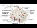 Bones: Structure and Types
 Chondrosarcoma vs Enchondroma: Bone Pathology with Dr. Andrew Rosenberg
 Identification of General Histology Slides
 Osteoblasts and Osteoclasts
 MICROSCOPIC STRUCTURES OF COMPACT BONE (WEDGE OF BONE)
 Picture tests in histology of the respiratory system
 CARTILAGE - Histology, Types, Functions
 Shotgun Histology Dense Bone
 Bone remodeling and repair
 Structure Of Bone Tissue - Bone Structure Anatomy - Components Of Bones
 Normal Bone Histology & Embryology 101 with Dr. Andrew Rosenberg
 Histology Bone marrow smear : Shotgun Histology
 Functions of Osteoblasts & Osteocytes | Organization of the Osteon
 Osteogenesis- Bone formation
 Bone tissue histology.avi
 Histology Helper - Bone & Cartilage Histology
 Bone Cells
 Soft Tissue & Bone Tumor Pathology Unknown Cases (Yale) (see video description for digital slides)
 Histology of Brain tissue ( CNS ) :Shotgun Histology