Histology of Spongy Bone : Shotgun Histology
►𝐉𝐨𝐢𝐧 𝐓𝐡𝐢𝐬 𝐂𝐡𝐚𝐧𝐧𝐞𝐥 𝐓𝐨 𝐆𝐞𝐭 𝐀𝐜𝐜𝐞𝐬𝐬 𝐓𝐨 𝐏𝐞𝐫𝐤𝐬 :- https://bit.ly/2RQHvTN
►𝐃𝐨𝐰𝐧𝐥𝐨𝐚𝐝 𝐭𝐡𝐞 𝐌𝐞𝐝𝐯𝐢𝐳𝐳 𝐚𝐩𝐩 𝐮𝐬𝐢𝐧𝐠 𝐭𝐡𝐞 𝐛𝐞𝐥𝐨𝐰 𝐥𝐢𝐧𝐤 👇👇👇👇 𝐃𝐨𝐰𝐧𝐥𝐨𝐚𝐝 👇👇👇👇
►𝐀𝐧𝐝𝐫𝐨𝐢𝐝 :- https://bit.ly/3ansFKq
📌𝐅𝐨𝐥𝐥𝐨𝐰 𝐨𝐧 𝐈𝐧𝐬𝐭𝐚𝐠𝐫𝐚𝐦 :-
https://www.instagram.com/drgbhanuprakash
Histology of Bone ( Spongy bone): Shotgun Histology
Types of the bone marrow
-------------------------------------------
Bone marrow lacks the rigidity of the surrounding bone. Instead, it is a jelly-like substance that fills the cavity left by the trabecular network of bone. Bone marrow accounts for about 4 – 5% of the total body weight of an individual. Although it can be considered a “light-weight” system, the bone marrow does a lot of heavy lifting, as it is responsible for producing platelets, lymphocytes, erythrocytes, granulocytes, and monocytes.
Marrow has two principal functions; one is to produce blood cells and the other is to store fat. As a result, there are two types of marrow found in the body:
the highly vascular red marrow which is haematopoietically active,
and the fat rich yellow marrow that has significantly less haematopoietic centres and more adipocytes.
Red bone marrow
==============
Clusters of haematopoietic cells known as haematopoietic islands are widely distributed throughout the loose connective tissue network observed in red marrow. These islands are found next to relatively large, yet thin walled, sinusoids that also communicate with nutrient vessels of the bone. The sinusoids are situated at a central part of a roundabout circulation such that the nutrient arteries that leave the nutrient canals to supply the bones anastomose in the bone marrow and subsequently terminate in arterioles that coalesce to form the sinusoids. The sinusoids then drain to significantly larger veins that form nutrient veins, which then leave the bone via the same nutrient canals that the arteries enter by.
Red marrow is most abundant in all skeletal structures from intrauterine life up until around the 5th year of life. As time progresses, red marrow is restricted to the central flat bones (i.e. cranial bones, clavicle, sternum, ribs, scapula, vertebrae, and pelvis) and the proximal ends of the proximal long bones of the upper and lower limbs.
The supporting substance that supports the haematopoietic and adipocyte cells in the marrow is made up of reticulin. This is a fine type III collagen that is produced by mesenchyme derived reticular cells (fibroblast-like cells). Other housekeeping cells like macrophages exist in the stroma and facilitate haematopoiesis by phagocytosing cellular debris generated from the process.
Yellow bone marrow
=================
Depending on the age and haematological demand of an individual, the reticular cells become swollen as a result of increased lipid uptake. Subsequently, yellow marrow is formed. It contains mainly supportive connective tissue that provides scaffolding for the neurovascular structures that traverse the cavitation. There are also numerous adipocytes in addition to very few dormant haematopoietic clusters. These latent haematopoietic centres can be reactivated in the event of an increase demand for red blood cells.
#histologyofspongybone #spongybone #bonehistology #spongybonehistology #histology #shotgunhistology
Видео Histology of Spongy Bone : Shotgun Histology канала Dr.G Bhanu Prakash Animated Medical Videos
Показать
Комментарии отсутствуют
Информация о видео
6 мая 2019 г. 23:06:39
00:04:13
Другие видео канала
 Histology of Bone (cortical bone) : Shotgun Histology
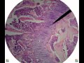 Bone tissue histology.avi
 MICROSCOPIC STRUCTURES OF COMPACT BONE (WEDGE OF BONE)
 Histology Bone marrow smear : Shotgun Histology
 Identifying Connective Tissue | Review and Practice
 Bone Biology: COMPACT BONE VS SPONGY BONE - EASY FAST REVIEW!!
 Histology of Spongy Bone
 Histology of the Spleen
 Types of Cartilage | Hyaline, Elastic, and Fibrocartilage
 Histology of Bone Tissue
 Shotgun Histology Endochondral Ossification
 Bone remodeling and repair
 Histology | Compact Bone (Osseous Tissue)
 Compact Bone Structure
 Intramembranous Ossification
 Hyaline Cartilage.mp4
 Identifying Muscle | Review and Practice
 Shotgun Histology Colon
 Identifying Epithelium | Review and Practice Questions
 How to interpret bone marrow aspirate and biopsy by Dr. Tejindar Singh, Oncquest Laboratories Ltd.