Picture tests in histology of the respiratory system
After completion of this video you will be able to:
Differentiate between
Trachea, bronchus and bronchiole
Type I and Type II alveolar cells
Alveolar duct and alveolar sac
Explain the role of the following respiratory passages structures:
Mucous, cilia, cartilage, smooth muscle, Clara cell, type I & II alveolar cells, alveolar macrophages, goblet cells, neuroendocrine cells, basal cells,
Identify: respiratory epithelium, basement membrane, cilia, goblet cell, basal cell, fibroblast, lymphocyte, dust cell
Presented and edited by Dr. Akram Jaffar, Ph.D.
This video and its channel are supported by "Human Anatomy Education" Page on Facebook http://www.facebook.com/AnatomyEducation
Subscribe to the channel to receive updates.
Feedback is highly appreciated from channel viewers
Some images, with gratitude, were cited in:
http://medicine.academic.ru/32539/metaplasia
http://www.columbia.edu/itc/hs/medical/sbpm_histology_old/lab/lab06_cartilage.html
https://www.slideshare.net/jrfisher78/bio12-respiratory-system-presentation
https://www.pinterest.com/wnethpoo/sem/
http://pathhsw5m54.ucsf.edu/case23/PNEC.html
http://ajplung.physiology.org/content/293/2/L259
https://commons.wikimedia.org/wiki/File:Clara_cell.jpg
Видео Picture tests in histology of the respiratory system канала Human Anatomy Education
Показать
Комментарии отсутствуют
Информация о видео
Другие видео канала
 Respiratory Histology – Histology | Lecturio
 Picture tests in histology of the cardiovascular system 1
 THE DEVELOPMENT OF THE LUNGS AND THE RESPIRATORY SYSTEM-HUMAN EMBRYOLOGY-DR ROSE JOSE MD
 Lungs: tissues and cells (preview) - Human Anatomy and Histology | Kenhub
 Picture tests in histology of the gastrointestinal system 1
 Histology of the lung explained by a lung pathologist. Part 2: bronchi
 Anatomy and physiology of the respiratory system
 Tissue Identification Practice
 Histology of the lung
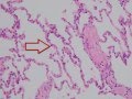 Histology of the Lung
 Picture test in histology of the endocrine glands
 LYMPHATIC HISTOLOGY
 Epithelium – Histology | Lecturio
 Shotgun Histology Trachea
 Picture tests in histology of the renal system 1
 Embryology of the Lungs (Easy to Understand)
 Functional Histology of the Respiratory System
 Respiratory | Mechanics of Breathing: Pressure Changes | Part 1
 Picture tests in histology of the cardiovascular system 2
 Respiratory bronchiole (histology of the lung)