ChIP Optimization: Tissue Processing Tips for ChIP
Loni Pickle, Life Technologies R&D Scientist, covers some key pointers for moving into tissue-based chromatin immunoprecipitation (ChIP) experiments.
Please refer to the MAGnify Chromatin Immunoprecipitation System manual for detailed information: http://tools.invitrogen.com/content/sfs/manuals/magnify_chip_man.pdf
Go to http://www.lifetechnologies.com/support to view our technical support resources, or to find out how to ask your question by email or phone.
AUDIO TRANSCRIPT:
Hey fellow ChIP researchers, and anyone new to the ChIP application, I'm Loni Pickle, fellow bench scientist, here to talk to you today about optimizing your ChIP, giving you some tips that I've learn from spectacular mentors over the years, spending a lot of time at the bench myself optimizing ChIP and also helping you guys out there in the field. So I wanted to bring you what I've learned here today. So what we're gonna talk about is tissue ChIP; we're gonna go through magnetic beads and how they're gonna make your life much easier when performing ChIP, and we're also gonna talk about shearing and shearing optimization."
Tissue Processing Using Magnetic Beads Shearing Optimization
Let's start with tissue processing for ChIP; a very important step in your ChIP application. When I moved from cells to tissue I was looking for a protocol that I could use. What I found out there it took too long, required me to purchase more expensive equipment, and it required a majority of my sample; which I wanted to use that sample for other purposes. So what I did was, we kinda developed our own protocol. Basically using a gradient of syringe needles; which made it quicker, made it extremely cheap, and cut down the amount of material to much, much less; meaning I could use that material for other purposes.
The important things that will be needed for this procedure are:
- 1 ml syringes
- 18 and 21 gauge needles
- 1 X PBS
- Petri Dishes
- Standard ChIP Reagents (formaldehyde, Lysis Buffer, and Glycine)
The five basic steps we are going to go over today are:
- Weighing
- Mincing
- Mashing
- Homogenizing with gradient needles
- Cross-linking
Step One: Weighing:
We are going to start by weighing between 50 to 1,000 milligrams of fresh or frozen tissue. You are going to want to put a Petri dish on ice, make sure everything is kept cold. You are going to add 250 ul of 1 X PBS to your Petri dish that has been placed on ice. You're gonna add between 50 to 1,000 milligrams of fresh or frozen tissue onto the Petri dish in order to do the second step which is mincing with a razor blade; so add your tissue.
Step Two: Mincing:
First remove any necrotic or fatty tissue from your tissue sample. Then you are going to mince the tissue using two razor blades into less than 1 mm cubed pieces. And all of these of course is done on ice and in 250 ul of 1 X PBS. So you are going to want to cut your tissue into less than 1 millimeter cubed pieces using the two razor blades. So make sure you finely dice the tissue, because the next steps we will be using syringes. We added 250 ul of ice cold PBS, but for more tissue than that, add 250 ul of ice cold PBS per 50 milligrams of tissue up to the maximum of 2 ml.
Step Three: Mashing:
We're going to transfer our tissue into 50 ml conical tube and mash the tissue in order to go through for homogenization. So you take a 2 ml pipette, and while the sample is still on ice collect your sample that has been in the media. Transfer it to an ice cold 50 ml conical tube. Use your pipette to mash the tissue as much as possible. We're going to further mash the tissue by adding a 1.5 ml 18 gauge needle onto your 1 ml syringe. Make sure you keep the sheathe on and mash the tissue further, to make it easier to homogenize in the next steps.
Step Four: Homogenizing with gradient needles:
Remove the sheathe and what we are going to do is we're going to pipette up and down 10 times to homogenize the tissue using your 18 gauge needle. It will go easier as you go along. If you happen to get a clog, all you do is remove it, and then expel it back through. Once you've homogenized 10 times using the 18 gauge needle you're going to quickly move to your 21 gauge needle; and with this needle you are going to homogenize and pipette up and down 20 times, and of course keeping the sample on ice as you're doing this. Once you've reached 20 times and the sample has been fully homogenized, and you can move on to formaldehyde cross-linking; which is step five.
Step Five: Cross-linking:
So after you've already homogenized your tissue, you're going to transfer your homogenized tissue to a brand new 1.5 ml tube at room temperature. The 50 milligrams of tissue is already in 250 ul of PBS so we're going to add an additional 200 ul of room temperature PBS to the sample. Which means, for every 50 milligrams of tissue we're going to have a final volume of 450 ul of PBS.
For more information visit http://owl.li/cPD2x
Видео ChIP Optimization: Tissue Processing Tips for ChIP канала Thermo Fisher Scientific
Показать
Комментарии отсутствуют
Информация о видео
22 ноября 2011 г. 3:02:54
00:07:28
Другие видео канала
 ChIP Optimization: Chromatin Shearing in ChIP
 How to use Dynabeads® for immunoprecipitation
 How to use the DynaMag™-5 Magnet.
 How to Prepare a Single-Cell Suspension from Mouse Brain Tissue
 Pathology tissue processing explained in 90 seconds
 Chromatin Immunoprecipitation (chip assay)
 Flow Cytometry Protocol for Staining Membrane Associated Proteins
 RNA extraction demonstration
 User Stories: LC-MS/MS Technology for the Clinical Laboratory, Belfast
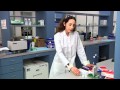 ChIP Optimization: Using Magnetic Beads in ChIP
 Tips for Handling and Storing Tissue Samples Prior to DNA Extraction
 5 Minute Guide to Human Organ-on-Chip Studies: Cells, Tissues, Organs and Multi-Organ Experiments
 Tissue Homogenization Video | Biovision, Inc.
 Plate-Based ELISA vs Invitrogen Dynabeads- Based ELISA
 Return to life with our COVID-19 Rapid PCR Test
 Tissue preparation
![[Tutorial] Cleaning coverslips using manual procedure | Mass Photometry](https://i.ytimg.com/vi/urPMpJ0x45o/default.jpg) [Tutorial] Cleaning coverslips using manual procedure | Mass Photometry
[Tutorial] Cleaning coverslips using manual procedure | Mass Photometry Bio-engineered scaffolding for skin
 Flow Basics 2.1: The Basic Staining Protocol
 Overview of Cup Horn Sonication