Femoral Triangle - Boundaries and Contents | Anatomy Tutorial
#anatomy #femoral #thigh
https://www.instagram.com/anatomy.knowledge/
link for donation: https://paypal.me/studentlamedicina?locale.x=en_US
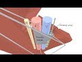
The femoral triangle (or Scarpa's triangle) is an anatomical region of the upper third of the thigh.
The femoral triangle is bounded:
• superiorly by the inguinal ligament.
• medially by the medial border of the adductor longus muscle
• laterally by the medial border of the sartorius muscle.
The following structures are contained within the femoral triangle (from lateral to medial):
Lateral cutaneous nerve of thigh crosses the lateral angle of the triangle.
Femoral nerve - The nerve enters the femoral triangle by passing beneath the inguinal ligament and after a short course of about 4 cm in the thigh, the nerve is divided into anterior and posterior divisions. The saphenous nerve is a brach from the posterior division of the femoral nerve. After its origin the saphenous nerve approaches the apex of the femoral triangle.
Femoral artery emerges from the base of the femoral triangle at the midpoint between the anterior superior iliac spine and the pubic symphysis of the pelvic bone and exits through the apex of the triangle into the adductor canal. Arising from the upper part of the femoral artery we have: the superficial circumflex iliac artery, the superficial epigastric artery and superficial and deep external pudendal arteries. The profunda femoris artery also arises from the femoral artery at the level of femoral triangle. One of the branches of profunda femoris is the lateral femoral circumflex artery which passes between the two divisions of femoral nerve.
Anteriorly to the femoral artery runs down the femoral branch of genitofemoral nerve.
The femoral vein lies medial to the femoral artery at the base of the triangle but as it approaches the apex of the triangle, it lies posteromedially to the femoral atery. It receives drainage from the grater saphenous vein which in turn receives drainage from the superficial circumflex iliac vein, superficial epigastric vein and superficial and deep external pudendal veins.
Deep inguinal limph nodes lies medial to the upper part of the femoral vein.
Видео Femoral Triangle - Boundaries and Contents | Anatomy Tutorial канала Anatomy Knowledge
https://www.instagram.com/anatomy.knowledge/
link for donation: https://paypal.me/studentlamedicina?locale.x=en_US
The femoral triangle (or Scarpa's triangle) is an anatomical region of the upper third of the thigh.
The femoral triangle is bounded:
• superiorly by the inguinal ligament.
• medially by the medial border of the adductor longus muscle
• laterally by the medial border of the sartorius muscle.
The following structures are contained within the femoral triangle (from lateral to medial):
Lateral cutaneous nerve of thigh crosses the lateral angle of the triangle.
Femoral nerve - The nerve enters the femoral triangle by passing beneath the inguinal ligament and after a short course of about 4 cm in the thigh, the nerve is divided into anterior and posterior divisions. The saphenous nerve is a brach from the posterior division of the femoral nerve. After its origin the saphenous nerve approaches the apex of the femoral triangle.
Femoral artery emerges from the base of the femoral triangle at the midpoint between the anterior superior iliac spine and the pubic symphysis of the pelvic bone and exits through the apex of the triangle into the adductor canal. Arising from the upper part of the femoral artery we have: the superficial circumflex iliac artery, the superficial epigastric artery and superficial and deep external pudendal arteries. The profunda femoris artery also arises from the femoral artery at the level of femoral triangle. One of the branches of profunda femoris is the lateral femoral circumflex artery which passes between the two divisions of femoral nerve.
Anteriorly to the femoral artery runs down the femoral branch of genitofemoral nerve.
The femoral vein lies medial to the femoral artery at the base of the triangle but as it approaches the apex of the triangle, it lies posteromedially to the femoral atery. It receives drainage from the grater saphenous vein which in turn receives drainage from the superficial circumflex iliac vein, superficial epigastric vein and superficial and deep external pudendal veins.
Deep inguinal limph nodes lies medial to the upper part of the femoral vein.
Видео Femoral Triangle - Boundaries and Contents | Anatomy Tutorial канала Anatomy Knowledge
Показать
Комментарии отсутствуют
Информация о видео
Другие видео канала
 3D Tour of the Femoral Triangle
 Femoral Artery and its branches - Anatomy tutorial
 Popliteal Fossa | Boundaries & Contents | Anatomy Tutorial
 Great Saphenous Vein & Small Saphenous Vein - Venous drainage of lower limb
 Sciatica - causes, symptoms, diagnosis, treatment, pathology
 Suprahyoid & Infrahyoid muscles of the neck | Anatomy Tutorial
 The Infratemporal Fossa - Boundaries & Contents | Anatomy Tutorial
 The Popliteal Artery branches & genicular anastomosis
 Obturator Nerve - Anatomy Tutorial
 Hypoglossal Nerve | Course & Branches | Anatomy Tutorial
 The Femoral Triangle under 5 mins! - Anatomy
 3D Tour of the Femoral Canal
 Anatomy Front of the thigh ( Femoral Triangle ) - Part 4 ( Dr.G.Bhanu Prakash )
 Facial Artery - Origin, course, branches | Anatomy Tutorial
 Clinical Anatomy - Knee
 Inguinal canal
 Muscles of the Hip and Thigh - Human Anatomy | Kenhub
 Cubital fossa anatomy - Boundaries, contents, and clinical anatomy
 Anatomy - Subclavian artery branches
 Ascending & Descending Tracts | Spinal Cord cross section Neuroanatomy