Neuroradiology board review 3 case 7
Neuroradiology board review. This lecture is geared towards the ABR core exam for residents, but it would be useful for review for the ABR certifying exam or certificate of added qualification (CAQ) exam for neuroradiology.
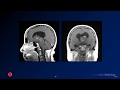
This case shows
expansion of the sella on a noncontrast CT, suggesting a long-standing mass. The MRI shows an underlying mass which has areas of hypointensity on T2 and intrinsic hyperintensity on T1 weighted imaging. There is minimal if any enhancement on post-contrast imaging.
The diagnosis is:
pituitary adenoma (with hemorrhage)
Pituitary adenomas are common masses of the sella, with about half being non-functional and half hormone secreting. The most common hormone secreted is prolactin, followed by growth hormone. Adenomas can hemorrhage, causing variable imaging appearance.
The primary differential consideration is Rathke cleft cyst, which is more commonly midline, less likely to have blood products and septations, and may have a characteristic peripheral nodule. You can read more about differentiating adenomas and Rathke cleft cysts here:
http://www.ajnr.org/content/36/10/1866
Check out this video and additional content on http://www.learnneuroradiology.com
Видео Neuroradiology board review 3 case 7 канала LearnNeuroradiology
Показать
Комментарии отсутствуют
Информация о видео
Другие видео канала
 Neuroradiology board review 3 case 1
 Neuroradiology board review 3 case 8
 Quarantine University- Imaging of Pituitary Gland and supraselllar region
 Power Foods for the Brain | Neal Barnard | TEDxBismarck
 Imaging of Sella I - DRE 5 - Dr Mamdouh Mahfouz
 Intracranial infections - 2 - Diffuse Infections
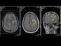 ADC Positive Multiple Sclerosis - Demyelination MRI Case Review
 Millennials in Medicine: Doctors of the Future | Daniel Wozniczka | TEDxNorthwesternU
 Neuroradiology review - brain gyral anatomy
 Cases in Radiology: Episode 1 (neuroradiology, CT, MRI)
 Radiology Review Unknowns | The Advanced EM Boot Camp - Maureen McCollough, MD, MPH
 Neuroradiology spotters -Dr M Venkatesh
 Neuroradiology Board Review - Brain Tumors - Case 18
 The most important lesson from 83,000 brain scans | Daniel Amen | TEDxOrangeCoast
 TMT: NEURORADIOLOGY: Short Cases Series 3
 Neuroradiology Board Review - Brain Tumors - Case 15
 Pineal Region Tumors: Radiologic-Pathologic Correlation
 e-Radiology Learning | Neuroradiology Pearls and Pitfalls (1 of 4)
 Neuroradiology Board Review - Brain Tumors - Case 13
 Intracranial infections - 1 - General principles