Neuroradiology board review 3 case 8
Neuroradiology board review. This lecture is geared towards the ABR core exam for residents, but it would be useful for review for the ABR certifying exam or certificate of added qualification (CAQ) exam for neuroradiology.
This case shows
a teenager with T2 hyperintense mass in the left posterior fossa resulting in mass effect on the 4th ventricle. On post-contrast imaging there is scattered hazy enhancement throughout portions of the mass.
The diagnosis is:
pilocytic astrocytoma
Pilocytic astrocytomas are the most common brain tumors in children. They most commonly occur in the posterior fossa and are often characterized by a cystic mass with a nodular enhancing area. They are relatively benign tumors with a good 10 year survival.
When you encounter a cyst with a nodule, there is a relatively short differential diagnosis which includes:
Pilocytic astrocytoma
Ganglioglioma
Hemangioblastoma (has blood vessels/flow voids)
Plemomorphic xanthoastrocytoma - PXA
Check out this video and additional content on http://www.learnneuroradiology.com
Видео Neuroradiology board review 3 case 8 канала LearnNeuroradiology
Показать
Комментарии отсутствуют
Информация о видео
Другие видео канала
 Neuroradiology board review 3 case 9
 Neuroradiology spotters -Dr M Venkatesh
 After watching this, your brain will not be the same | Lara Boyd | TEDxVancouver
 Neuroradiology Board Review - Brain Tumors - Case 13
 Imaging brain tumors - 1 - Introduction and classification
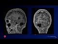 Neuroradiology Board Review - Brain Tumors - Case 6
 Neuroradiology review - brain gyral anatomy
 Your brain hallucinates your conscious reality | Anil Seth
 Brain magic | Keith Barry
 Tumors of the Central Nervous System - CRASH! Medical Review Series
 Posterior fossa neoplasms
 IDKD Refresher Series - Pediatric Neuroradiology
 Intracranial infections - 1 - General principles
 Neuroradiology board review 3 part 20 - multiple choice review
 Cases in Radiology: Episode 1 (neuroradiology, CT, MRI)
 Report skills Part 2: Report framework and content
 Neuroradiology Board Review - Brain Tumors - Case 20 - Summary
 TMT: NEURORADIOLOGY: Short Cases Series 3
 GLIOBLASTOMA MULTIFORME (GBM) -RADIOPATH CORRELATION
 PEDIATRIC POSTERIOR FOSSA NEOPLASMS | AMITKUMAR CHOUDHARI | MR IMAGING | MEDULLOBLASTOMA | GLIOMA