Intracranial infections - 1 - General principles
Intracranial infections are a common consideration in neuroradiology. Patients often present with altered mental status, often with a fever or systemic symptoms. The radiologist needs to know how to proceed in those instances, both in terms of what types of imaging to perform and how imaging findings might relate to the systemic findings.
This lecture is the first in a series of 5 about intracranial infection and covers general principles behind imaging the patient with suspected intracranial infection. The subsequent 4 videos cover additional more specific considerations.
The two main imaging techniques used in imaging intracranial infection are CT and MRI. CT is a rapid screening test which can detect edema, hemorrhage, midline shift, and prominent masses. It is available in the vast majority of hospitals (at least in the US, but also many abroad), and doesn't require much compliance from the patient. It also requires no screening for metallic implants and other MRI considerations.
MRI is much better at evaluating intracranial infection however. Key sequences include DWI, FLAIR, T2, and T1 postcontrast. DWI is ideal for seeing pus and infarct. FLAIR is ideal for imaging edema. T2 can give you some unique clues, as a few diseases such as lymphoma, tuberculosis, and abscess can be T2 dark. Many infections also cause breakdown of the blood brain barrier, which shows up as enhancement on postcontrast MRI.
Lumbar puncture and echocardiogram are the two main systemic tests which may help you determine if an intracranial infection is present. LP directly assesses for infections agents and inflammatory cells in the CSF, while echocardiogram indirectly assesses for bacterial endocarditis, which can spread to the CSF through emboli.
The level of this lecture is appropriate for radiology residents, radiology fellows, and trainees in other specialties who have an interest in neuroradiology or may see patients with CNS infections.
Check out this video and additional content on http://www.learnneuroradiology.com
Видео Intracranial infections - 1 - General principles канала LearnNeuroradiology
Показать
Комментарии отсутствуют
Информация о видео
Другие видео канала
 Intracranial infections - 2 - Diffuse Infections
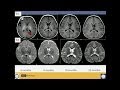 Neuroimaging of Pediatric Disease
 Multiple sclerosis – white spots and red flags - part 1 - Making a diagnosis
 How to read a CT angiogram (CTA) of the Head and Neck
 Imaging CNS autoimmune and inflammatory disease - 2 - Encephalitis
 Intracranial infections - 3 - Focal Infections
 MSK Radiology Cases - Spotters Set 6 : Quiz and Discussion (LIVE)
 Neuroradiology Part 1
 Imaging CNS autoimmune and inflammatory disease - 1 - Introduction/Demyelinating disease
 NEURORADIOLOGY: CNS Infections - I | DEEPAK PATKAR | Tuberculomas vs Cysticercosis
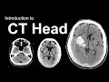 Introduction to CT Head: Approach and Principles
 Intracranial infections - 4 - Immunocompromise
 Neuroradiology board review 3 case 4
 Imaging of pediatric intracranial infections - DRE 7 - Dr Mamdouh Mahfouz
 Emergency Spinal Radiological Assessment
 Overview of Congenital Heart Disease CT/MRI
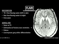 MRI Sequences
 MRI basic (level 1), for beginner
 Isolation tutorial: Neuroradiology #3 with Frank Gaillard
 MRA (magnetic resonance angiogram) head radiology search pattern