Imaging CNS autoimmune and inflammatory disease - 2 - Encephalitis
This is the second lecture in a case based review of imaging of the brain and spine for autoimmune and inflammatory conditions. We will cover the MRI findings of some of the common conditions and some potential pitfalls and mimics.
This second lecture covers causes of encephalitis, including inflammatory and autoimmune etiology. This section also covers infectious encephalitis, which is the most common mimic.
1:05 Limbic encephalitis
Limbic encephalitis is an immune mediated encephalitis often associated with neoplasm and a variety of circulating antibodies. It most commonly is manifested as bilateral (asymmetric or symmetric) temporal lobe FLAIR/T2 abnormalities. Enhancement is variable but less common than seen in herpes encephalitis.
3:33 Chronic limbic encephalitis
Over time, chronic limbic encephalitis can result in temporal lobe sclerosis and atrophy. Volumetric analysis of the brain and temporal lobes may be useful in highlighting the differences over time.
5:39 Lupus encephalitis
Lupus is a systemic autoimmune condition which can be associated with intracranial findings, including encephalitis, vasculitis, and posterior reversible encephalopathy syndrome (PRES). Vessel imaging may be normal, as it tends to affect the small vessels of the brain.
9:04 Viral encephalitis
Viral encephalitis has a nonspecific imaging appearance with patchy white matter FLAIR/T2 abnormalities, often involving the temporal lobes. There are a number of causes of viral encephalitis, including herpes, west nile virus, St. Louis encephalitis, and others.
11:06 Herpes encephalitis
Herpes encephalitis is the most dreaded of the encephalitis causes because it has high morbidity and mortality. Compared to nonspecific viral and inflammatory encephalitis, it tends to have more enhancement and more DWI abnormality. Patients suspected of having herpes encephalitis should be started empirically on treatment immediately to improve the prognosis.
12:59 Summary and Conclusion
The level of this lecture is appropriate for radiology residents, radiology fellows, and trainees in other specialties, such as neurology, who have an interest in neuroradiology or may see patients with CNS demyelinating or inflammatory conditions.
NOTE: I'm disabling comments on this video because I'm getting too much spam about herpes.
Check out this video and additional content on http://www.learnneuroradiology.com
#neuroradiology #radiology #encephalitis
Видео Imaging CNS autoimmune and inflammatory disease - 2 - Encephalitis канала LearnNeuroradiology
Показать
Комментарии отсутствуют
Информация о видео
Другие видео канала
 Imaging CNS autoimmune and inflammatory disease - 1 - Introduction/Demyelinating disease
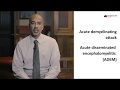 Autoimmune Encephalitis - an Overview
 Imaging CNS autoimmune and inflammatory disease – Part 3 - Masslike inflammatory disease
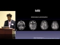 Posterior Reversible Encephalopathy Syndrome (PRES): Who, What When? by Tommy T. Thomas, MD, PhD
 Encephalitis (“Brain Inflammation”) Signs and Symptoms (& Why They Occur)
 Vascular Imaging of the Head and Neck - Case A
 Brain MRI - Seizure search pattern
 Vascular Imaging of the Head and Neck - Case C
 Imaging CNS autoimmune and inflammatory disease - 4 - Spine inflammatory disease
 Encephalitis - My Brain: My Story 2019 - Mallory's Story
 CNS Vasculitis: Diagnostic Update
 Multiple sclerosis – white spots and red flags - part 1 - Making a diagnosis
 Imaging CNS autoimmune and inflammatory disease - 5 - Amyloid related disease
 Hydrocephalus And the Disorders of CSF Circulation
 Encephalitis - My Brain: My Story 2019 - Hannah's Story
 HSV Encephalitis - Pathogenesis, Clinical Presentation, and Diagnosis
 How to read a CT angiogram (CTA) of the Head and Neck
 Noncontrast MRI cervical spine search pattern
 The #1 CAUSE Of Autoimmune Disease & How To PREVENT IT! | Mark Hyman
 Epilepsy Imaging