Histology of trachea - Shotgun Histology
Histology of trachea - Shotgun Histology
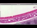
The trachea is a wide flexible tube, the lumen of which is kept open by 20 tracheal cartilages, which are C-shaped rings of hyaline cartilage. The gaps between the rings of cartilage are filled by the trachealis muscle - a bundle of smooth muscle, and fibroelastic tissue. Together these hold the lumen of the trachea open, but allow flexibility during inspiration and expiration.
The respiratory mucosa and submucosa are adapted to warm and moisten the air, and to trap particles in mucous.
Mucosa and sub-mucosa of Trachea
The respiratory mucosa is made up of the epithelium and supporting lamina propria). The epithelium is tall columnar pseudostratified with cilia and goblet cells. The supporting lamina propria underneath the epithelium contains elastin, that plays a role in the elastic recoil of the trachea during inspiration and expiration, together with blood vessels that warm the air.
The sub-mucosa contains glands which are mixed sero-mucous glands. The watery secretions from the serous glands humidify the inspired air. The mucous, together with mucous from the goblet cells traps particles from the air which are transported upwards towards the pharynx by the cilia on the epithlium. This helps to keep the lungs free of particles and bacteria.
#histologyoftrachea #tracheahistology #shotgunhistology #tracheahistologyvideo #tracheahistologyslide #tracheashotgunhistology
Видео Histology of trachea - Shotgun Histology канала Dr.G Bhanu Prakash Animated Medical Videos
Показать
Комментарии отсутствуют
Информация о видео
12 мая 2019 г. 0:34:49
00:05:15
Другие видео канала
 Histology of the trachea
 Shotgun Histology Parotid Gland
 Shotgun Histology Lung
 (R)evolutionary Medicine: Rachel Abrams at TEDxSantaCruz
 Trachea Histology
 Shotgun Histology Spinal Cord
 Dr Paul Lee - Treating Diabetes and Obesity Through Brown Fat
 Histology of Bone
 Trachea: cells, tissues, histology slides (preview) - Human histology | Kenhub
 Histology: Blood Cells
 USMLE Step 1 Buzzwords (Part 1)
 Histology of the lung
 Embryology | Development of the Respiratory System
 MED STUDENT VLOG (USMLE STEP 1 EXPERIENCE PART 2)
 Trachea | Extent | Dimensions | Structure | Relations | Clinical Anatomy
 Larynx - Membranes, ligaments and muscles - Human Anatomy | Kenhub
 Picture tests in histology of the respiratory system
 Pathophysiology of COPD
 Histologie Ösophagus - Mikroskopische Anatomie - AMBOSS Video
 Shotgun Histology Liver