Anatomy of the Skull : Norma Verticalis and Norma Occipitalis
Join this channel to get access to perks:
https://www.youtube.com/channel/UCG5TBPANNSiKf1Dp-R5Dibg/join
Follow on Instagram :- https://www.instagram.com/drgbhanuprakash
Anatomy of the Skull: Norma Verticalis and Norma Occipitalis
Norma Verticalis.—When viewed from above the outline presented varies greatly in different skulls; in some, it is more or less oval, in others more nearly circular. The surface is traversed by three sutures, viz.: (1) the coronal sutures, nearly transverse is direction, between the frontal and parietals; (2) the sagittal sutures, medially placed, between the parietal bones, and deeply serrated in its anterior two-thirds; and (3) the upper part of the lambdoidal suture, between the parietals and the occipital. The point of junction of the sagittal and coronal suture is named the bregma, that of the sagittal and lambdoid sutures, the lambda; they indicate respectively the positions of the anterior and posterior fontanelles in the fetal skull. On either side of the sagittal suture are the parietal eminence and parietal foramen—the latter, however, is frequently absent on one or both sides. The skull is often somewhat flattened in the neighborhood of the parietal foramina, and the term obelion is applied to that point of the sagittal suture which is on a level with the foramina. In front is the glabella, and on its lateral aspects are the superciliary arches, and above these the frontal eminences. Immediately above the glabella may be seen the remains of the frontal suture; in a small percentage of skulls, this suture persists and extends along the middle line to the bregma. Passing backward and upward from the zygomatic processes of the frontal bone are the temporal lines, which mark the upper limits of the temporal fossæ. The zygomatic arches may or may not be seen projecting beyond the anterior portions of these lines.
Norma Occipitalis.—When viewed from behind the cranium presents a more or less circular outline. In the middle line is the posterior part of the sagittal suture connecting the parietal bones; extending downward and lateralward from the hinder end of the sagittal suture is the deeply serrated lambdoidal suture joining the parietals to the occipital and continuous below with the parietomastoid and occipitomastoid sutures; it frequently contains one or more sutural bones. Near the middle of the occipital squama is the external occipital protuberance or inion, and extending lateralward from it on either side is the superior nuchal line, and above this the faintly marked highest nuchal line. The part of the squama above the inion and highest lines is named the planum occipitale, and is covered by the Occipitalis muscle; the part below is termed the planum nuchale, and is divided by the median nuchal line which runs downward and forward from the inion to the foramen magnum; this ridge gives attachment to the ligamentum nuchæ. The muscles attached to the planum nuchale are enumerated on p. 130. Below and in front are the mastoid processes, convex laterally and grooved medially by the mastoid notches. In or near the occipitomastoid suture is the mastoid foramen for the passage of the mastoid emissary vein.
#normaverticalis #normaoccipitalis #skullanatomy #skull #osteology #anatomy #anatomyoftheskull #anatomyofcranialbones #cranialbones #skullbones #occipitalbone #anatomy #osteology
Видео Anatomy of the Skull : Norma Verticalis and Norma Occipitalis канала Dr.G Bhanu Prakash Animated Medical Videos
Показать
Комментарии отсутствуют
Информация о видео
5 сентября 2020 г. 18:30:14
00:11:58
Другие видео канала
 Anatomy of the Skull : Norma Frontalis
 FMGE AUG 31st obstetrics and gynecology Recall Questions with Dr. Ramya sree
 Chest pain: how to distinguish between cardiac and noncardiac causes. Dr.Magesh.T MD( USA) MRCP(UK)
 Axillary nerve Anatomy : Origin, Course, Branches, innervation and clinical anatomy | Medvizz
 Diabetes mellitus series ( Part 1 ) - Types of Diabetes, Causes, Risk Factors, and Symptoms
 True Story of a successful medical PG Aspirant ( Must watch )
 Norma Occipitalis | The study posterior aspect of skull bones
 Rapid Revision Obstetrics for FMGE
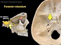 The Skull
 Anatomy - Cranial Nerves Overview
 FMGE AUG 31st Medicine Recall Questions with Dr. Rajesh Gubba
 Temporal bone
 NORMA OCCIPITALIS
 Norma Frontalis | Study of skull from front or anterior
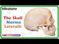 Anatomy of the Skull : Norma Lateralis
 Skull foramina
 Practical Practice that's Practically Priceless! Bones and structures of the skeletal system.
 Fetal Skull in Hindi (हिंदी)| Practical Explanation | Nursing Lecture
 SKELETAL SYSTEM ANATOMY: Inferior aspect of the human skull
 Anatomy of head & neck 10 ( Skull , part 10 , norma basalis externa ) , by Dr. Wahdan