How to Interpret a Chest X-Ray (Lesson 6 - Diaphragm and Pleura)
A review of how to diagnose a pneumothorax, various forms of pleural effusion, other forms of pleural disease, and pneumoperitoneum. A differential diagnosis for each of these findings is discussed as well.
Video includes the following images (among others):
Right pleural effusion, downloaded from Radiopaedia.org, originally provided by Roberto Schubert
Pleural plaques, downloaded from Radiopaedia.org, originally provided by Jeremy Jones
Presumed mesothelioma, downloaded from Radiopaedia.org, originally provided by Jeremy Jones
Pneumoperitoneum, downloaded from Radiopaedia.org, originally provided by Henry Knipe
Chilaiditi's sign, downloaded from Radiopaedia.org, originally provided by Hani Salam.
Sources for other images may include Wikimedia Commons, radiologypics.com, and Jose Caceres' wonderful radiology blog: Caceres Corner (http://blog.myesr.org/category/caceres-corner/)
Видео How to Interpret a Chest X-Ray (Lesson 6 - Diaphragm and Pleura) канала Strong Medicine
Показать
Комментарии отсутствуют
Информация о видео
Другие видео канала
 How to Interpret a Chest X-Ray (Lesson 7 - Diffuse Lung Processes)
 How to Interpret a Chest X-Ray (Lesson 5 - Cardiac Silhouette and Mediastinum)
 Diaphragms and Pleural Effusion - How to Read a Chest X-Ray (Part 8) - MEDZCOOL
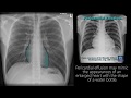 Radiograph Tutorial: Chest X-ray / CXR | Radiology Nation
 Intro to EKG Interpretation - Chamber Enlargement
 Pneumonia: Types, Classification, Symptoms & Management – Respiratory Medicine | Lecturio
 Pneumothorax - Spontaneous, Tension & Traumatic
 How to Interpret a Chest X-Ray (Lesson 8 - Focal Lung Processes)
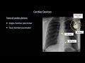 How to Interpret a Chest X-Ray (Lesson 9 - Atelectasis, Lines, Tubes, Devices, and Surgeries)
 How to Interpret a Chest X-Ray (Lesson 10 - Self Assessment): Part 1
 Dr Bhatia discussing on PLEURAL EFFUSION in #LastMinuteRevisionPointDiscussionSeries
 Chest X-ray: Cases 1
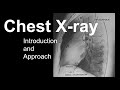 Chest X-ray: Introduction and Approach
 How to Interpret a Chest X-Ray (Lesson 1 - An Introduction)
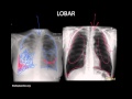 Pneumonia: Imaging
 02 CXR: DRS ABCDE Method
 LEARN to Read a Chest Xray in 5 minutes!
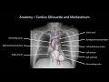 How to Interpret a Chest X-Ray (Lesson 2 - A Systematic Method and Anatomy)
 Pleural Effusion Explained by Prometheus Lionhart, MD
 Chest x-ray - Pneumothorax or no pneumothorax