How to Interpret a Chest X-Ray (Lesson 7 - Diffuse Lung Processes)
An explanation of alveolar vs. interstitial opacities, including cardiogenic and non-cardiogenic pulmonary edema, and the 3 types of interstitial patterns (reticular, nodular, and reticulonodular). Examples provided of air bronchograms, peribroncial cuffing, Kerley A and B lines, and cephalization. Etiologies of low lung volumes and hyperinflation are also discussed.
Video includes the following image (among others):
Cephalization, downloaded from Radiopaedia.org, originally posted by Charlie Chia-Tsong Hsu.
Sources for other images may include Wikimedia Commons, radiologypics.com, and Jose Caceras' wonderful radiology blog: Caceres Corner (http://blog.myesr.org/category/caceres-corner/)
Видео How to Interpret a Chest X-Ray (Lesson 7 - Diffuse Lung Processes) канала Strong Medicine
Показать
Комментарии отсутствуют
Информация о видео
Другие видео канала
 Pulmonary Exam Demo (Strong Exam)
 SOAP Presentations - 2 Examples
 I Walked Through the World's Largest Heart
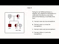 Clinical Vignettes on Hemostasis (Less Common Scenarios) - Hemostasis: Lesson 14
 Clinical Vignettes on Hemostasis (Common Scenarios) - Hemostasis: Lesson 13
 A Doctor's 100 Pet Peeves About Hospital Medicine (100-51)
 How to Provide Effective Inpatient Consultation
 EAP Presentations - 2 Examples
 The Threshold Model of Clinical Decision-Making (Strong Diagnosis)
 Viscosity and Turbulence
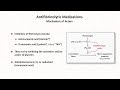 Prothrombotic Medications: Hemostasis - Lesson 7
 Acute Hypertension (Rapid Response Calls)
 A Patient's General Appearance (Strong Exam)
 Rapid Response Calls (Intern Crash Course)
 Acute Somnolence (Rapid Response Calls)
 Acute Respiratory Distress (Rapid Response Calls)
 Gastroparesis
 The woman who faked a brain tumor to avoid prison - UPDATE
 Underappreciated Diseases - An Introduction
 Acute Chest Pain (Rapid Response Calls)
 The Pulmonary Exam / Lung Sounds (Strong Exam)