ACL Tears Radiological Evaluation - Everything You Need To Know - Dr. Nabil Ebraheim
Dr. Ebraheim’s educational animated video describes radiological evaluations of Anterior Cruciate Ligament (ACL) tears.
Follow me on twitter:
https://twitter.com/#!/DrEbraheim_UTMC
The Anterior Cruciate Ligament (ACL) is a strong band of tissue in the center of the knee that prevents anterior translation of the tibia on the femur. The mechanism of injury to the ACL is usually a noncontact, pivoting injury. As the ACL tears, the patient feels a “POP” and deep pain in the knee. The patient usually feels immediate swelling and the knee is usually filled with blood. This occurs due to tear of the middle genicular artery. The patient usually feels immediate swelling and the knee is usually filled with blood. This occurs due to tear of the middle genicular artery. On examination, the patient will have a quadriceps avoidance gait.
The Lachman’s Test is the most sensitive test to diagnose tear of the ACL. During the Lachman’s Test, the knee should be bent to about 20-30 degrees. One hand should be used to stabilize the femur and the other hand should pull the tibia anteriorly and posteriorly, against the femur. If the ACL is ruptured, the ACL will be lax and the examination will feel softer with no end point. The tibia can be pulled forward more than normal (anterior translation).
On x-ray, you can see Anterior Cruciate Ligament (ACL) avulsion. Avulsion of a piece of bone from the tibial eminence anteriorly. Avulsion of a piece of bone can be seen on the AP and lateral x-rays. An ACL avulsion fracture may be an isolated injury or it may sometimes be associated with other injuries and fractures such as tibial plateau fractures. Tibial spine fractures in children mimics ACL avulsion fractures in adults. An axial view MRI is used to evaluate the proximal ACL. This “Empty Notch Sign” or “Empty Wall Sign” indicates a torn ACL. The “Empty Notch Sign” means that there is an avulsion of the proximal femoral attachment of the ACL. A sagittal view MRI will show disruption of the ACL fibers. An MRI may show nonvisible and noncontinuous fibers. MRI will show the proximal ACL fibers to be dangled and the distal fibers to be dropped due to complete disruption of the ACL. ACL tears may cause bone bruises laterally on the middle of the femoral condyle and on the posterior aspect of the tibia laterally. In order to determine if a bone bruise is lateral, look at the fibula. The fibula is lateral. Check for the fibula on MRI. The pattern of bone marrow edema seen of lateral view and AP view MRI that will indicate an ACL tear. An impaction fracture of the lateral femoral sulcus terminalis. Suspect impaction fracture and ACL tears when the lateral sulcus depth is greater than 2mm. A fibular head avulsion, or arcuate sign, is a horizontally oriented fracture of the fibular styloid process, the attachment site of the arcuate ligament complex. It can be associated with injury of the ACL, the PCL, and the posterolateral corner (probably more associated with the PCL than the ACL). A fibular head avulsion fracture will indicate that the posterolateral corner is involved. The arcuate sign should be recognized as a significant injury. Sometimes the avulsed piece of bone is too small and the injury can be missed. Failure to diagnose a fibular head avulsion may result in failure of future fixation of the cruciate ligaments, because the posterolateral corner instability was not diagnosed and treated properly.
Acute ACL injury may be associated with lateral meniscus injury. Chronic ACL injury may be associated with medial meniscus injury. In the ACL deficient knee, the posterior horn of the medial meniscus provides the secondary restraint to the anterior tibial translation. This added stress on the posterior horn of the medial meniscus may cause the medial meniscus to become injured.
Видео ACL Tears Radiological Evaluation - Everything You Need To Know - Dr. Nabil Ebraheim канала nabil ebraheim
Показать
Комментарии отсутствуют
Информация о видео
Другие видео канала
 Pivot Shift Test ACL Tear - Everything You Need To Know - Dr. Nabil Ebraheim
 Systematic Interpretation of Knee MRI: How I do it
 What is ACL Surgery?
 Clinical Anatomy - Knee
 What are ACL Injuries - Learn the Symptoms, Risk Factors, Why it Gets Torn, and More!
 ACL, PCL, & Quadriceps - Everything You Need To Know - Dr. Nabil Ebraheim
 Arthritis Of The Knee - Everything You Need To Know - Dr. Nabil Ebraheim
 My ACL Surgery Recovery Journey As a Physiotherapist
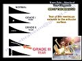 Knee Pain, Meniscal Tear Diagnosis & MRI - Everything You Need To Know - Dr. Nabil Ebraheim
 Knee Plica and Knee pain - Everything You Need To Know - Dr. Nabil Ebraheim
 ACL Surgery - 3D Reconstruction
 Anatomy Of The Knee - Everything You Need To Know - Dr. Nabil Ebraheim
 Medial Collateral Ligament Injuries - Everything You Need To Know - Dr. Nabil Ebraheim
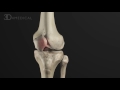 Anterior Cruciate Ligament: Pathology and Management | Animated Tutorial
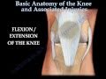 Knee injury ,Injuries - Everything You Need To Know - Dr. Nabil Ebraheim
 Anatomy Of The Psoas & Iliacus Muscles - Everything You Need To Know - Dr. Nabil Ebraheim
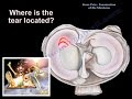 Knee Pain , Meniscus tear - Everything You Need To Know - Dr. Nabil Ebraheim
 Anterior Cruciate Ligament (ACL) Injuries
 Anterior Cruciate Ligament (ACL) Injuries
 How to Read Knee MRI of ACL Tear | Anterior Cruciate Ligament Pain | Knee Surgery | Minneapolis, MN