How to Perform a Full Volume Acquisition on a Philips EPIQ Ultrasound
http://www.usa.philips.com/healthcare-education-resources/education-training/ultrasound-education-instructional-guides
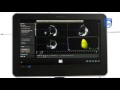
This video offers a step-by-step guide on how to acquire a full volume dataset on a Philips EPIQ ultrasound system. In this example, the X5-1 transducer is used with the apical 4 chamber view. Data can be acquired from different views depending on your target anatomy.
Full volume acquisition is used when larger volume of data is needed, usually including the whole heart as when performing an ejection fraction in QLAB.
Tips:
- Make sure there is a strong ECG signal with regular R-R intervals.
- Remember to adjust depth, focus, and iScan for optimal results.
What's next? Need the 3D volume of the left ventricle? Check out this step-by-step video for 3D Quantification Advanced (3DQA) [link to video].
Видео How to Perform a Full Volume Acquisition on a Philips EPIQ Ultrasound канала Philips Healthcare
Показать
Комментарии отсутствуют
Информация о видео
Другие видео канала
 How To Perform 3D Quantification Using Philips QLAB 3DQA Application
 Philips Cardiovascular Ultrasound: 3D Auto RV
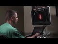 Philips TrueVue, TouchVue, MPR Touch, GlassVue, aReveal
 Philips OB/GYN Ultrasound: How to acquire a 3D or 4D obstetric volume
 Philips Cardiovascular Ultrasound: AutoStrain RV
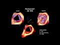 3D Echocardiography Acquisition, Cropping, and Case Examples
 How to Perform a 4D Acquisition on a Philips EPIQ Ultrasound System
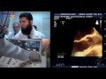 How to perform 3D-TEE exam "a step by step approach"
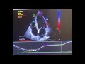 How to do strain echocardiogram ❤️✨
 LOGIQ F8 Getting Started F8 DOC1471565 | GE Healthcare
 Philips OB/GYN Ultrasound: How to perform a 3D GYN volume
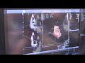 Philips Epiq 7 vs GE Vivid E95 Premium Cardiac Ultrasound System Comparison
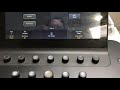 3rd In Series How to operate Philips Epiq ultrasound machine
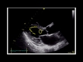 Estimating Ejection Fraction with Point of Care Echo
 Carotid Protocol (Esther Collado, RN, RVI)
 Philips Cardiovascular Ultrasound: AutoStrain LA
 Ultrasound training PHILIPS ClearVue 650
 Philips Ultrasound: ElastQ Imaging for liver assessment
 Knobology 1-A Optimising ECHO Images Dr Hafeesh Fazulu 1st July 2021 Class 1 - Part 1
 How to assess the mitral valve area using 3D Echocardiography