How To Perform 3D Quantification Using Philips QLAB 3DQA Application
http://www.usa.philips.com/healthcare-education-resources/education-training/ultrasound-education-instructional-guides
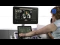
This video is a step-by-step guide on how to use 3D Quantification Advanced (3DQA) on a Philips EPIQ ultrasound system. 3DQA is a semi-automated border detection tool designed for use with 3D cardiac images in QLAB. 3DQA can be used to analyze full volume datasets for a true 3D volume of the left ventricle. Results measure regional and global function, including ejection fraction and LV synchronicity. 3DQA provides a global volume of the heart with no apical foreshortening or geometrical assumption and has been validated again cardiac MR.
Once a Full Volume dataset is acquired, 3DQA is used to set reference points in the apical 2 and for chamber views at the mitral annulus and apex of the left ventricle for the end systole and end diastole frames. 3DQA will then automatically detect the endometrial border and generate a true 3D volume of the left ventricle.
Want more? Subscribe to this playlist and visit our education page for handy step-by-step QuickGuides! http://www.usa.philips.com/healthcare-education-resources/education-training/ultrasound-education-instructional-guides
Видео How To Perform 3D Quantification Using Philips QLAB 3DQA Application канала Philips Healthcare
Показать
Комментарии отсутствуют
Информация о видео
Другие видео канала
 How to Perform a Full Volume Acquisition on a Philips EPIQ Ultrasound
 A Guideline for Best Practices in 3-D Imaging: Dr. Roberto Lang at iMAGINE
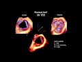 3D Echocardiography Acquisition, Cropping, and Case Examples
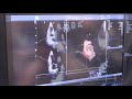 Philips Epiq 7 vs GE Vivid E95 Premium Cardiac Ultrasound System Comparison
 Fetal Echocardiography: Protocol and Technique
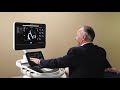 Philips Cardiovascular Ultrasound: AutoStrain RV
 Heart model - Frank Kemme, Sr. Clinical Application Specialist Ultrasound, Philips Healthcare
 Philips OB/GYN Ultrasound: How to perform a 3D GYN volume
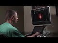 Philips TrueVue, TouchVue, MPR Touch, GlassVue, aReveal
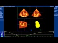 How to use LV quantification software of Q-Lab 9
 Ultrasound EPIQ new era in Ultrasound, Intelligence , Performance and Design
 A novel method to measure Mitral Valve Area "The MVN method".
 3D Echocardiography: Latest Advances and Applications (ROBERTO M. LANG, MD)
 Perform a Lumify Exam: Philips Lumify product training (5 of 11)
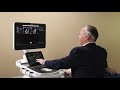 Philips Cardiovascular Ultrasound: AutoStrain LA
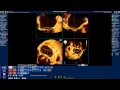 How to use Mitral Valve Quantification (MVQ) software of Q-Lab
 Philips Cardiovascular Ultrasound: 3D Auto RV
 How to measure the 3D-Vena Contracta Area (3D-VCA) for mitral regurgitation
 Strain Echocardiography by speckle tracking and tissue Doppler -Part I:technique
 How to perform 3D-TTE exam "a step by step approach"