MRI Anatomy of ankle ligaments: Syndesmosis
SUPPORT MY CHANNEL HERE: https://www.patreon.com/agten
Become a faster Radiologist - buy my book on Amazon: https://www.amazon.com/dp/1074826930/
In this video, part 3 of my ankle ligament series, I cover the syndesmosis and all other ligaments of the distal tibiofibular joint. Including the anteroinferior tibiofibular ligament, the interosseous tibiofibular ligament, the posteroinferior tibiofibular ligament, the interosseous membrane, the intermalleolar ligament, and Bassets ligament. In addition, I show you important anatomic landmarks such as the Wagstaff Le-Fort tubercle, Chaput's tubercle, Volkmann's tubercule and the incisura fibularis of the tibia.
This anatomy is very important for the assessment of MRI with regards to high ankle sprains, which are not rare, especially in athletes.
#anklesprain
#anatomy
#MRI
References:
https://www.ncbi.nlm.nih.gov/pubmed/31289899
Please subscribe to my channel and also check out my patreon page: https://www.patreon.com/agten
Patreon is an online system, where you can support me on a more personal level with a tiny donation in exchange for small benefits, as listed on my page. It is a great way to engage with me and learn together. Every month I post patreon-only videos over on my patreon page.
Thanks for watching and keep learning!
Видео MRI Anatomy of ankle ligaments: Syndesmosis канала Dr Christoph Agten
Показать
Комментарии отсутствуют
Информация о видео
Другие видео канала
 MRI Anatomy of lateral ankle ligaments
 Syndesmosis Injury - When Is It Necessary To Refer For Surgery
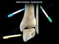 Ankle Fractures and the Syndesmosis - Everything You Need To Know - Dr. Nabil Ebraheim
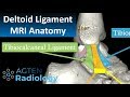 MRI Anatomy of Ankle Ligaments: Deltoid Ligament
 Radiology Boards Prep - More MSK Cases!
 Knee Examination Inspection & Palpation - Everything You Need To Know - Dr. Nabil Ebraheim
 Imaging of the Tibiofibular Syndesmosis and High Ankle Sprain
 Osteochondral Defects of the Ankle | Dr. Helder Pereira (Portugal)
 Ankle Anatomy Animated Tutorial
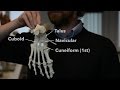 Foot and ankle bones
 Systematic Interpretation of Ankle MRI: How I do it
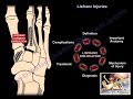 Lisfranc Injuries - Everything You Need To Know - Dr. Nabil Ebraheim
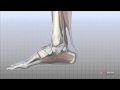 Foot Anatomy Animated Tutorial
 Clinical Anatomy - Lower Limb (Bones), Inguinal ligament, Hip, Knee and ankle Joints
 MRI Elbow Anatomy 1920 x 1080
 Sacroiliitis or not? with Prof. Hermann - Part 3
 Syndesmotic Injuries Of The Ankle - Everything You Need To Know - Dr. Nabil Ebraheim
 MRI of the iliopsoas bursitis and its pitfalls
 MRI of anterior ankle impingement
 How to learn Radiology - Top 10