Ankle Fractures and the Syndesmosis - Everything You Need To Know - Dr. Nabil Ebraheim
Dr. Ebraheim’s educational animated video describing fractures of the ankle fractures - syndesmotic injury.
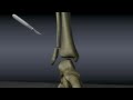
how do we know if we have a syndesmotic injury? By getting the intra-operative stress exam, external rotation of the talus within the ankle mortise, this test determine if syndesmotic instability is present, you will do that test after fixation of the other fractures.
The abduction external rotation of the talus will try to displace fibula from the incisura fibularis, the talus will move laterally and displaces the fibula.
The ankle will show a valgus talar tilt or increase in the medial clear space.
Before you do syndesmotic reduction and fixation, it is important to restore the length and rotation of the fibula.
When instability is present, you have to do syndesmotic screw fixation.
How do you know if there is instability? Always have a high index of suspicion.
Syndesmotic fixation is more required when the fibular fracture is high and there is a deltoid ligament injury.
Be skeptical about some of the statements such as fixation is not typically required when the fibular fracture is within 4.5 cm from the joint because that is not true.
Just remember: Weber C is commonly associated with syndesmotic injury.
So we get the stress views and look at certain measurements to determine if the syndesmosis is injured or not.
At 1 cm above the joint we will measure the tibulofibular overlap which will be decreased if there is a syndesmotic injury.
We also measure the tibiofibular clear space which will be more than 5 mm if there is a syndesmotic injury.
Then we look at the medial clear space which will be increased, normally it should be less than 4 mm.
Some people believe that the instability of the ankle appears more in the AP plain.
The medial clear space can be increased preoperatively due to injury to the deltoid ligament.
This is used to differentiate between supination – external rotation stage II and stage IV injuries.
The medial clear space can be helpful intraoperatively after fixation of the fibula to diagnose syndesmotic injury on stress view radiographs.
Syndesmosis fixation techniques:
- You must restore the length and rotation of the fibula, which is not good enough by itself.
- An Accurate reduction of the syndesmosis is required and direct inspection of the syndesmotic reduction is helpful, and this should be supported by x-rays.
- Check for widening.
- Check for the chenton’s line, dime sign, and that will be done after reduction and after using the reduction clamp.
- This is the time to get an AP view and lateral view radiographs, and you assess before you place your screws.
Try to use multiple techniques to check on the syndesmosis injury, one of them is the external rotation view the intraoperative one.
The other one is the cotton test, get a hook and pull on the fibula and see the movement.
The third one is direct inspection of the syndesmosis, make sure the crural fascia may be intact and covering a major syndesmotic injury.
After that we go to the technique:
1- You dorsiflex the ankle.
2- Directly inspect and reduce the fibula.
3- Use reduction clamp.
4- Get x-rays to prove that the syndesmosis is reduced and then you put the screws, about 2-4 cm above the joint, with an angle of 20° to 30° posteriorly to anteriorly.
Do not use lag screws and do not over compress the syndesmosis with the position of the talus in planter flexion, although a lot of people think it is not possible.
Screws are really controversial ad no consensus about them.
But there are a few important points about the screws:
1- The 4.5 mm are not used a lot nowadays.
2- When the widening is bad you are going to use more screws and more cortices, the more the better.
3- When you put the screws proximally and you don’t aim anteriorly you may miss the tibia.
4- Make sure when you go from cortex 1 to 2 and 3 in the tibia that you don’t miss cortex number 3 in the tibia.
5- Try to elevate the ankle a little bit so your hand will be allowed to do some anterior direction of the screws, so the screws will be angled a little bit.
6- Occasionally I cross the screws, so will be one direct straight forward and the other one will be oblique.
7- Screw removal: it’s controversial but you will not remove the screws before 3 months.
What are the problems with the syndesmosis?
• Missing the injury: Reading the x-ray, I use the 5 mm for reading the x-rays, whatever it is in the medial clear space or tibiofibular clear space as my mark, 5 mm is abnormal.
• Malreduction of the syndesmosis: I want to make sure the fibula is anatomically reduced to the incisura before inserting the syndesmotic screws; I want to make sure and get an x-ray to check the talus both in the AP and lateral planes.
Dr. Ebraheim is an orthopedic surgeon at the University of Toledo Medical Center.
Видео Ankle Fractures and the Syndesmosis - Everything You Need To Know - Dr. Nabil Ebraheim канала nabil ebraheim
Показать
Комментарии отсутствуют
Информация о видео
Другие видео канала
 Ankle Fractures
 Imaging of the Tibiofibular Syndesmosis and High Ankle Sprain
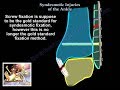 Syndesmotic Injuries Of The Ankle - Everything You Need To Know - Dr. Nabil Ebraheim
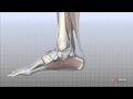 Foot Anatomy Animated Tutorial
 Malleolar ankle fractures - evaluation (OTA lecture series III l12a)
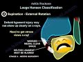 Ankle Fractures - Everything You Need To Know - Dr. Nabil Ebraheim
 Syndesmosis or High Ankle Sprain Treatment
 How does a plate and screws help a broken bone heal?
 NERVE INJURY IN THE UPPER EXTREMITY- Everything You Need To Know - Dr. Nabil Ebraheim
 Tibial Pilon Fracture - Everything You Need To Know - Dr. Nabil Ebraheim
 Syndesmotic injuries - screws vs dynamic fixation and other controversies
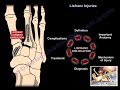 Lisfranc Injuries - Everything You Need To Know - Dr. Nabil Ebraheim
 Ankle Pain, ankle ligaments sprain - Everything You Need To Know - Dr. Nabil Ebraheim
 Fibula
 Tibia - Intraarticular Fracture - Large External Fixator- Ankle-bridging Delta Frame
 Calcific Tendonitis of the shoulder - Everything You Need To Know - Dr. Nabil Ebraheim
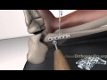 Operative treatment of ankle fractures
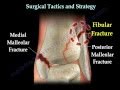 Ankle fracture / Fractures and its repair- Everything You Need To Know - Dr. Nabil Ebraheim
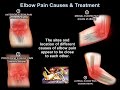 Elbow Pain Causes & Treatment - Everything You Need To Know - Dr. Nabil Ebraheim
 Ankle Fractures , Special Situations - Everything You Need To Know - Dr. Nabil Ebraheim