Anatomy Of The Flexor Hallucis Longus Muscle - Everything You Need To Know - Dr. Nabil Ebraheim
Dr. Ebraheim’s educational animated video describes the anatomy of the Flexor Hallucis Longus Muscle.
The Flexor Hallucis Longus Muscle is one of the deep muscles of the posterior compartment of the leg. The FHL muscle spans down the calf to the side of the ankle into the foot. The muscle fibers have the lowest origin in the leg and come fibers are almost at the ankle.
Origin and insertion: the FHL muscle arises from the posterior surface on the inferior two-thirds of the fibula. The FHL is the largest and strongest deep muscle of the leg’s posterior section.
There are two sesamoid bones at the level of the MCP joint, one tibular and one fibular, that act like pulleys for the flexor tendons and are embedded into the tendons of the Flexor Hallucis Brevis Muscle. Sesamoid are important to the big toe region by absorbing weight-bearing pressure and reducing friction on the metatarsal head.
The Flexor Hallucis Longus tendon has a long course from its muscle origin to its insertion. The tendon is vulnerable to wear, tear, friction and inflammation at certain anatomic locations. At the ankle, the FHL is located posterior and lateral to the FDL, between the posterior talar processes. The tendon lies most lateral in the medial ankle compartment of the posterior talus. The flexor hallucis tendonitis usually occurs in ballet dancing and other activities that require excessive plantar flexion of the ankle. As the tendons curve under the medial malleolus and talus, they begin to converge and eventually cross. The FHL is crossed deep to the flexor digitorum longus tendon into the medial compartment of the foot. This point of crossing is called the Knot of Henry. At this point, the fibrous slip connects the flexor digitorum longus with the Flexor Hallucis Longus at the Knot of Henry and the tendon sheath communicates allowing for spreading of inflammation. Because of the intersection of the two tendons, transaction of the digitorum proximal to the Knot of Henry to correct the tibialis posterior dysfunction could result in retention of the function of the hallux and the lesser digits.
Become a friend on facebook:
http://www.facebook.com/drebraheim
Follow me on twitter:
https://twitter.com/#!/DrEbraheim_UTMC
Background music provided as a free download from YouTube Audio Library.
Song Title: Every Step
Видео Anatomy Of The Flexor Hallucis Longus Muscle - Everything You Need To Know - Dr. Nabil Ebraheim канала nabil ebraheim
Показать
Комментарии отсутствуют
Информация о видео
Другие видео канала
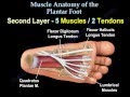 Muscle Anatomy Of The Plantar Foot - Everything You Need To Know - Dr. Nabil Ebraheim
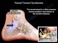 Tarsal Tunnel Syndrome - Everything You Need To Know - Dr. Nabil Ebraheim
![Flexor Hallucis Longus PAIN [FHL Tendonitis BEST Treatment 2021!]](https://i.ytimg.com/vi/C6Glw_3YBsw/default.jpg) Flexor Hallucis Longus PAIN [FHL Tendonitis BEST Treatment 2021!]
Flexor Hallucis Longus PAIN [FHL Tendonitis BEST Treatment 2021!]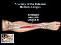 Anatomy Of The Extensor Hallucis Longus Muscle - Everything You Need To Know - Dr. Nabil Ebraheim
 Anatomy Of The Tibialis Anterior Muscle - Everything You Need To Know - Dr. Nabil Ebraheim
 FHL Tendon Transfer Using the DX Button and Tension-Slide Technique
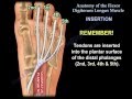 Anatomy Of The Flexor Digitorum Longus Muscle - Everything You Need To Know - Dr. Nabil Ebraheim
 Anatomy Of The Extensor Digitorum Longus Muscle - Everything You Need To Know - Dr. Nabil Ebraheim
 Flexor Hallucis Longus Pain – Anatomy, Diagnoses, and Treatment
 Flexor Hallucis Longus FHL Tendon Transfer
 Ankle Pain - Everything You Need To Know - Dr. Nabil Ebraheim
 Brachial plexus
 Flexor Hallucis Longus & Flexor Digitorum Longus (FHL & FDL) Static Manual Release
 Anatomy Of The Gastrocnemius Muscle - Everything You Need To Know - Dr. Nabil Ebraheim
 A Review Of Acetabular Fractures - Everything You Need To Know - Dr. Nabil Ebraheim
 Bringing the Foot Back To Life: Restoring the Extensor Hallucis Brevis Muscle.
 Tibialis Posterior Tendinopathy | Tendinitis | Dysfunction | Pain (Exercises, Rehab, Strengthening)
 Ankle Pain Complete Overview - Everything You Need To Know - Dr. Nabil Ebraheim
 Functions of the flexor digitorum longus muscle (preview) - 3D Human Anatomy | Kenhub
 Anatomy Of The Adductor Magnus Muscle - Everything You Need To Know - Dr. Nabil Ebraheim