Anatomy Of The Gastrocnemius Muscle - Everything You Need To Know - Dr. Nabil Ebraheim
Dr. Ebraheim’s educational animated video describes the anatomy of the gastrocnemius muscle in the leg.
Follow me on twitter:
https://twitter.com/#!/DrEbraheim_UTMC
The gastrocnemius muscle is a part of the superficial flexors of the leg. There are three superficial flexor muscles in the leg: gastrocnemius, soleus, and plantaris. The gastrocnemius muscle arises from the
femur and it crosses the knee joint and the ankle joint. The gastrocnemius muscle has two heads medial and lateral head. The origin has two heads: the lateral head arises from the lateral surface of the later condyle of the femur. The medial head of the gastrocnemius comes from the posterior surface of the femur above the medial femoral condyle. The medial head is larger and extends lower than the lateral head of the gastrocnemius. The two heads of the gastrocnemius muscle unite together near the middle of the leg. The two heads are separated by a furrow in which the sural nerve and the small saphenous vein are present. The two bellies of the gastrocnemius muscle end in a tendon which joins the tendon of the soleus muscle to form together the tendoachilles or the tendoncalcaneus, which inserts into the middle third of the posterior surface of the calcaneus. The gastrocnemius muscle is innervated by the tibial nerve (S1-S2) and each head of the muscle has its own branch. The tibial nerve passes behind the gastrocnemius and soleus muscles through the fibrous arch of the soleus. Each head of the gastrocnemius muscle is supplied by the sural branch of the popliteal artery. Together with the soleus muscle, the gastrocnemius muscle is a powerful plantar flexor of the ankle. Because the gastrocnemius muscle crosses the knee joint, it also flexes the knee. The gastrocnemius muscle is involved in running and jumping (it is a fast movement muscle). The gastrocnemius muscle contains white, type II fast twitch muscle fibers in contrast to the soleus muscle, which contains type I slow twitch muscle fibers. The flexor muscle groups are two compartments (superficial and deep). The gastrocnemius muscle is part of the superficial flexor compartment of the leg. These are the fasciotomy incisions for the two incision technique. It opens all four compartments including the superficial flexor groups. The gastrocnemius muscle is part of the boundaries of the popliteal fossa. The popliteal fossa is bounded by the biceps femoris superiorly and laterally, as well as the semitendinosus and semimembranosus superiorly and medially. The lower part of the space is formed by the two heads of the gastrocnemius muscle. The Baker’s cyst lies between the semimembranosus and the medial head of the gastrocnemius (cross-section used in ultrasound). A Baker’s cyst is commonly caused by knee arthritis or a meniscal tear. The cyst is connected to the knee joint through a valvular opening. Knee effusion from intra-articular pathology allows the fluid to go through the valve to the cyst in one direction. You approach this posteromedial fracture fragment of the tibial plateau through an incision between the medial gastrocnemius and the semimembranosus muscles. This fragment should have its own fixation (antiglide plate) to buttress this fracture fragment. Improved ankle dorsiflexion wit knee flexion means gastrocnemius muscle tightness or contracture. When you have the same ankle dorsiflexion with the knee in flexion or in extension, this means there is Achilles tightness. In gastrocnemius tightness or contracture, the treatment is gastrocnemius recession which can be helpful in diabetic foot ulcers. In gastrocnemius tightness or contracture, the treatment is gastrocnemius recession which can be helpful in diabetic foot ulcers. Medial gastrocnemius rotation flap can be used for proximal tibia open fractures and large soft tissue defects around the knee. The gastrocnemius muscle extends the distal fragment (the apex is posteriorly). The hamstrings and the extensor muscles cause shortening of the femur. The adductor magnus causes varus of the distal fragment.
Видео Anatomy Of The Gastrocnemius Muscle - Everything You Need To Know - Dr. Nabil Ebraheim канала nabil ebraheim
Показать
Комментарии отсутствуют
Информация о видео
Другие видео канала
 Anatomy of the calf (posterior leg)
 Gastrocnemius and Soleus Manual Static Release (Trigger Point Release)
 Anatomy Of The Soleus Muscle - Everything You Need To Know - Dr. Nabil Ebraheim
 Calf Tear, Strain, or Pain? Absolute Best Self Treatment and Exercises.
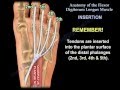 Anatomy Of The Flexor Digitorum Longus Muscle - Everything You Need To Know - Dr. Nabil Ebraheim
 The Most Scientific Way to Train CALVES (Science Explained)
 Do This One Thing Right & Your Calf Pain/Strain/Tear Will Heal Fast-See NEW Product at End of Video
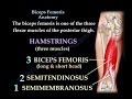 Biceps Femoris Anatomy, Hamstrings - Everything You Need To Know - Dr. Nabil Ebraheim
 Calf Strains, Tendinitis, & Achilles Tears | The Anatomy of the Calves
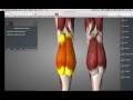 Muscles of lower leg:ankle
 Anatomy Of The Peroneal Muscles In The Lower Leg - Everything You Need To Know - Dr. Nabil Ebraheim
 Arthritis Of The Fingers - Everything You Need To Know - Dr. Nabil Ebraheim
 Functions of the gastrocnemius muscle (preview) - 3D Human Anatomy | Kenhub
 Large hip muscles
 How to treat a Calf strain (Gastrocnemius/Soleus) using Kinesiology Tape
 Gluteal muscles (anatomy)
 How To FORCE YOUR CALVES To Grow With Smarter Training Methods
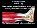 Anatomy Of The Lower Leg - Everything You Need To Know - Dr. Nabil Ebraheim
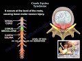 Cauda Equina Syndrome - Everything You Need To Know - Dr. Nabil
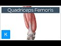 Quadriceps Femoris Muscle - Origin, Insertion and Function - Human Anatomy | Kenhub