Complications Of Talar Neck Fractures - Everything You Need To Know - Dr. Nabil Ebraheim
Dr. Ebraheim’s educational animated video describes complications of talar neck fractures.
Follow me on twitter:
https://twitter.com/#!/DrEbraheim_UTMC
Find me on Instagram @OrthoInitiative
Complications of Talar Neck Fractures
There are multiple complications for talar neck fractures: arthritis, avascular necrosis (best diagnosed with x-rays), malunion, nonunion (occurs in about 5%). Subtalar arthritis is the most common complication of talar neck fractures. Subtalar arthritis occurs in 50-100% of cases (occurs more than AVN). Ankle joint arthritis, which is tibiotalar joint arthritis, occurs in about 1/3 of patients (33% of patients). Avascular necrosis means “death of a segment of a bone” due to interruption of the blood supply. In the case of talar neck fractures, there will be death of the body of the talus. Osteonecrosis does not usually involve the entire talar body. The artery of the tarsal canal is the dominant blood supply. The deltoid branch of the posterior tibial artery is the only remaining blood supply with displaced talar neck fractures. The deltoid branch of the tarsal cannel must be preserved during surgery. It supplies half of the medial talar body. The blood supply of the talar neck is very tenuous and when the talus fractures, this blood supply is susceptible to injury and disruption. The incidence of AVN correlates with the degree of displacement and the severity of the fracture. The extent of the initial fracture displacement is very important. The risk of AVN increases fracture displacement. If the blood supply is seriously affected, then that will increase the incidence of AVN. There are four types of talar neck fractures and the incidence of AVN varies in each type. The incidence of AVN is about 30% for all types. AVN occurs more with open fractures. Type I is a non-displaced fracture of the talar neck and has 10% incidence of AVN. Type II is a displaced fracture of the subtalar dislocation or subluxation and has about 50% AVN. Type III is a displaced fracture, and the talar body is subluxed or dislocated from the subtalar joint and also from the ankle joint, and the incidence of AVN is about 90%. Type IV is a displaced fracture with talar head subluxation from the talonavicular joint, and the body is extruded and has about 100% AVN. When AVN occurs, the condition becomes complicated and salvage procedure can be helpful. The status of the talar vascularity can be checked by the Hawkin’s sign. AVN is diagnosed on plain x-rays and by the absence of the Hawkin’s sign. The Hawkin’s sign is a subchondral osteopenia lucency seen at 6-8 weeks on the mortise view x-ray of the ankle of the dome of the talus. Look for this radiolucent line below the subchondral bone, which is more commonly seen on the medial side of the mortise view. The Hawkin’s sign is a good indication of intact vascularity with resorption of the subchondral bone following fractures of the talar neck. Presence of the Hawkin’s sign indicates that the talus is alive, and the prognosis is good. Absence of the Hawkin’s sign does not rule out an intact vascularity. In all patients that had the Hawkin’s sign, none of them developed avascular necrosis. The sensitivity is 100%, but the specificity is about 57%. No association has been shown between the timing of the fixation and the development of osteonecrosis. The fracture is usually reduced and fixed. The patient is followed up clinically and by x-rays for healing of the fracture and for the development of avascular necrosis. Once the fracture heals, begin weight bearing. Restricting weight bearing beyond that which is needed for healing of the fracture does not decrease the incidence of avascular necrosis. At 3-6 months postoperatively, AVN can bee seen on the plain x-ray as sclerosis. The MRI is sensitive for detecting AVN as it shows decreased signal onT1, but it does not guide the treatment. In MRI studies, titanium implants have better visualization than stainless steel implants. Varus malunion occurs in about 25-30% of cases. The patient will have varus and decreased subtalar motion, especially eversion. You will find that the patient ambulates on the lateral aspect of the foot. Varus malunion occurs due to medial comminution of the talus. It is a preventable complication. In talus fracture, it is important to restore the articular cartilage reduction, shape, and the axial alignment of the talus. Caution is suggested when compression fixation is used in the medial side of the talus, especially when there is a medial comminution, because this will lead to a varus deformity. The use of medial and lateral approaches will help to avoid this complication of varus malunion. The treatment of varus malunion is medial opening wedge osteotomy of the talar neck. Impingement of the dorsal surface of the talus on the distal tibia and decreased ankle dorsiflexion. This deformity can occur due to an isolated dorsal malunion.
Видео Complications Of Talar Neck Fractures - Everything You Need To Know - Dr. Nabil Ebraheim канала nabil ebraheim
Показать
Комментарии отсутствуют
Информация о видео
Другие видео канала
 Talus Fractures - Everything You Need To Know - Dr. Nabil Ebraheim
 General Trauma Management,the injured patient- Everything You Need To Know - Dr. Nabil Ebraheim
 Diabetic Ankle Fractures - Everything You Need To Know - Dr. Nabil Ebraheim
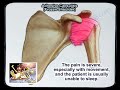 Adhesive Capsulitis Frozen Shoulder - Everything You Need To Know - Dr. Nabil Ebraheim
 Scaphoid Fractures - Everything You Need To Know - Dr. Nabil Ebraheim
 Foot Drop Peroneal Nerve Injury - Everything You Need To Know - Dr. Nabil Ebraheim
 A Review Of Acetabular Fractures - Everything You Need To Know - Dr. Nabil Ebraheim
 Ankle Fractures , Anatomical Considerations - Everything You Need To Know - Dr. Nabil Ebraheim
 Perilunate Instability & Dislocation - Everything You Need To Know - Dr. Nabil Ebraheim
 Talus, Neck - Fractures - Fixation
 Tibial Pilon Fracture - Everything You Need To Know - Dr. Nabil Ebraheim
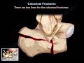 Calcaneal Fractures - Everything You Need To Know - Dr. Nabil Ebraheim
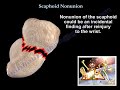 Scaphoid Nonunion - Everything You Need To Know - Dr. Nabil Ebraheim
 Ankle Fractures - Everything You Need To Know - Dr. Nabil Ebraheim
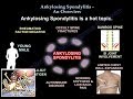 Ankylosing Spondylitis An Overview - Everything You Need To Know - Dr. Nabil Ebraheim
 Cervical Radiculopathy - Everything You Need To Know - Dr. Nabil Ebraheim
 Spinal cord injury ,Sacral Sparing - Everything You Need To Know - Dr. Nabil Ebraheim
 Acetabular Fracture Transverse Fracture - Everything You Need To Know - Dr. Nabil Ebraheim
 Ankle Fractures,and Posterior Malleolar Fracture - Everything You Need To Know - Dr. Nabil Ebraheim
 Fractures Of The Calcaneus - Everything You Need To Know - Dr. Nabil Ebraheim