Meckel's Diverticulum Made Easy
LIKE US ON FACEBOOK : fb.me/Medsimplified
BUY USING AFFILIATE LINKS :
AMAZON US--- https://goo.gl/XSJtTx
AMAZON India http://goo.gl/QsUhku
FLIPKART http://fkrt.it/Wiv8RNNNNN
FLIPKART MOBILE APP http://fkrt.it/Wiv8RNNNNN
A Meckel's diverticulum, a true congenital diverticulum, is a slight bulge in the small intestine present at birth and a vestigial remnant of the omphalomesenteric duct (also called the vitelline duct or yolk stalk). It is the most common malformation of the gastrointestinal tract and is present in approximately 2% of the population,[1] with males more frequently experiencing symptoms.
Meckel's diverticulum was first explained by Fabricius Hildanus in the sixteenth century and later named after Johann Friedrich Meckel, who described the embryological origin of this type of diverticulum in 1809
Meckel's diverticulum is located in the distal ileum, usually within 60–100 cm (2 feet) of the ileocecal valve. This blind segment or small pouch is about 3–6 cm long and may have a greater lumen diameter than that of the ileum.[4] It runs antimesenterically and has its own blood supply. It is a remnant of the connection from the yolk sac to the small intestine present during embryonic development. It is a true diverticulum, consisting of all 3 layers of the bowel wall which are mucosa, submucosa and muscularis propria.[5]
As the vitelline duct is made up of pluripotent cell lining, Meckel’s diverticulum may harbor abnormal tissues, containing embryonic remnants of other tissue types. Jejunal, duodenal mucosa or Brunner's tissue were each found in 2% of ectopic cases. Heterotopic rests of gastric mucosa and pancreatic tissue are seen in 60% and 6% of cases respectively. Heterotopic means the displacement of an organ from its normal anatomic location.[6] Inflammation of this Meckel's diverticulum may mimic appendicitis. Therefore during appendectomy, ileum should be checked for the presence of Meckel's diverticulum, if it is found to be present it should be removed along with appendix.
A memory aid is the rule of 2s:[7]
2% (of the population)
2 feet (proximal to the ileocecal valve)
2 inches (in length)
2 types of common ectopic tissue (gastric and pancreatic)
2 years is the most common age at clinical presentation
2:1 male:female ratioSymptoms[edit]
The majority of people with a Meckel's diverticulum are asymptomatic. An asymptomatic Meckel's diverticulum is called a silent Meckel's diverticulum.[9] If symptoms do occur, they typically appear before the age of two years.
The most common presenting symptom is painless rectal bleeding such as melaena-like black offensive stools, followed by intestinal obstruction, volvulus and intussusception. Occasionally, Meckel's diverticulitis may present with all the features of acute appendicitis. Also, severe pain in the epigastric region is experienced by the patient along with bloating in the epigastric and umbilical regions. At times, the symptoms are so painful that they may cause sleepless nights with acute pain felt in the foregut region, specifically in the epigastric and umbilical regions.
In most cases, bleeding occurs without warning and stops spontaneously. The symptoms can be extremely painful, often mistaken as just stomach pain resulting from not eating or constipation.
Diagnosis[edit]
Technetium-99m Pertechnetate Scan with a Meckel's Diverticulum.
A technetium-99m (99mTc) pertechnetate scan, also called Meckel scan, is the investigation of choice to diagnose Meckel's diverticula in children. This scan detects gastric mucosa; since approximately 50% of symptomatic Meckel's diverticula have ectopic gastric or pancreatic cells contained within them,[10] this is displayed as a spot on the scan distant from the stomach itself. In children, this scan is highly accurate and noninvasive, with 95% specificity and 85% sensitivity;[5] however, in adults the test is only 9% specific and 62% sensitive.[11]
Patients with these misplaced gastric cells may experience peptic ulcers as a consequence. Therefore, other tests such as colonoscopy and screenings for bleeding disorders should be performed, and angiography can assist in determining the location and severity of bleeding. Colonoscopy might be helpful to rule out other sources of bleeding but it is not used as an identification tool.
https://www.youtube.com/channel/UCOmrniWfKi-uCD6Oh6fqhgw
watch again
https://youtu.be/nQ8rZFhy_QA
-~-~~-~~~-~~-~-
CHECK OUT NEWEST VIDEO: "Nucleic acids - DNA and RNA structure "
https://www.youtube.com/watch?v=0lZRAShqft0
-~-~~-~~~-~~-~-
Видео Meckel's Diverticulum Made Easy канала MEDSimplified
Показать
Комментарии отсутствуют
Информация о видео
Другие видео канала
 Intussusception - causes, symptoms, diagnosis, treatment, pathology
 Meckel's Diverticulum With Clinical Correlations
 Meckel's Diverticulum made easy! | Embryology | Anatomy |
 Diverticulitis Signs & Symptoms (And Why They Occur)
 Diverticular Disease (diverticulitis) - Overview
 Meckel's Diverticulum (1-minute review)
 Hirschsprung disease (congenital aganglionic megacolon) - causes & symptoms
 Volvulus - causes, symptoms, diagnosis, treatment, pathology
 Meckel's Diverticulum : Anatomy
 Animated Gross anatomy of Appendix: Position, Blood supply, Venous drainage, Nerve supply, Histology
 Meckel's diverticulum - Pathology mini tutorial
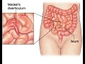 Meckel Diverticulum
 Bowel Obstruction - Causes and Pathophysiology
 SIGNS THAT YOU HAVE A LIVER DISEASE/ liver disease signs and symptoms
 Ulcerative colitis - causes, symptoms, diagnosis, treatment, pathology
 VITAMIN D DEFICIENCY/The Sunshine vitamin
 Acute appendicitis USMLE Step 1 : Etiology, Pathophysiology, Clinical Features, Diagnosis, Treatment
 Omphalocele and Gastroschisis
 Introduction to Direct and Indirect Inguinal Hernia
 Meckel's diverticulum by Dr A K Singh