Animated Gross anatomy of Appendix: Position, Blood supply, Venous drainage, Nerve supply, Histology
FOLLOW ON INSTAGRAM:- https://www.instagram.com/drgbhanuprakash/
Channel Memberships: https://www.youtube.com/channel/UCG5TBPANNSiKf1Dp-R5Dibg/join
Animated Gross anatomy of Appendix - Position, Blood supply, Venous drainage, Nerve supply and Histology
The cecum and appendix are part of the gastrointestinal tract. They are the most proximal part of the large intestine, located between the ileum (distal small bowel) and the ascending colon.
Having served as a site for cellulose digestion in our ancestors, the cecum now simply acts as a reservoir for chyme which it receives from the ileum. The appendix contains a large amount of lymphoid tissue, but has no vital functions in the human.
In this article, we shall look at the anatomy of the cecum and appendix – their anatomical structure and relations, neurovascular supply and lymphatic drainage.
Anatomical Structure and Relations
The cecum. Note the blind end inferiorly, and its continuity with the ascending colon superiorly.
The cecum. Note the blind end inferiorly, and its continuity with the ascending colon superiorly.
The cecum is the most proximal part of the large intestine and can be found in the right iliac fossa and suprapubic region. It lies slightly inferior to the iliocaecal junction, and can be palpated in the right iliac fossa if enlarged due to faeces, gas or malignancy.
The name is derived from the Latin word ‘caecus’, meaning ‘blind’, due to its blind-end inferiorly. Superiorly, however, the open end of the cecum is continuous with the ascending colon. Unlike the ascending colon above it, the cecum is intraperitoneal.
The appendix, also known as the vermiform (worm-shaped) appendix is a narrow, blind ended tube attached to the posteromedial end of the cecum. The position of the free-end of the appendix is highly variable, and can be categorised into seven main locations. The most common positions are retrocaecal and subileal. A simple way to remember the positions is by imagining the appendix as the hour hand of a clock:
Pre-ileal – Anterior to the terminal ileum – 1 o’clock.
Post-ileal – Posterior to the terminal ileum – 2 o’clock.
Sub-ileal – Parallel with the terminal ileum – 3 o’clock.
Pelvic – Descending over the pelvic brim – 5 o’clock.
Sub-caecal – Below the cecum – 6 o’clock.
Paracaecal – Alongside the lateral border of the cecum – 10 o’clock.
Retrocaecal – Behind the cecum – 11 o’clock.
Neurovascular Supply
-----------------------------------
The cecum is derived from the embryologic midgut – and therefore blood is supplied by a branch of the superior mesenteric artery. The branch is the ileocolic artery, which splits into anterior and posterior caecal arteries to supply the cecum. Venous drainage is provided by the ileocolic vein, which empties into the superior mesenteric vein.
The appendix receives its blood supply via the appendicular artery (derived from the ileocolic artery), and drains through the appendicular vein. Both are contained, along with lymphatic vessels and nerves, within the mesoappendix, a fold of mesentery which suspends the appendix from the terminal ileum.
The autonomic nervous system innervates the cecum and appendix. It achieves this by means of the ileocolic branch of the superior mesenteric plexus, which follows the same course as the ileocolic artery.
The cecum, appendix and ascending colon. Note the inferior position of the cecum in relation to the ileum.
Arterial supply to the cecum and appendix, via the ileocecal artery.
Lymphatic Drainage
-------------------------------
Lymph from the cecum and appendix ultimately drains into the upper and lower ileocolic lymph nodes, which surround the ileocolic artery.
However, lymph from the cecum travels via a number of intermediate mesenteric nodes, whereas that of the appendix travels via a single intermediate node in the mesoappendix.
Clinical Relevance: Appendicitis
---------------------------------------------------
Appendicitis is acute inflammation of the appendix, and is the most common cause for acute, severe abdominal pain. The abdomen is most tender at McBurney’s point – one third of the distance from the right anterior superior iliac spine to the umbilicus. This corresponds to the location of the base of the appendix.
The aetiology depends on age. In the young, it is mostly due to an increase in lymphoid tissue size, which occludes the lumen. From 30 years old onwards, it is more likely to be blocked due to faecal matter.
Initially, the appendix cannot drain, and so increases in size, stretching the visceral peritoneum. This causes a vague pain in the periumbilical region. As the appendix swells, it irritates the parietal peritoneum, and causes severe pain in the right lower quadrant.
If the appendix is not removed, it can become necrotic and rupture, resulting in peritonitis (inflammation of the peritoneum).
#anatomyofappendix #grossanatomyofappendix #appendix #appendixanatomy #anatomy #anatomyvideos
Видео Animated Gross anatomy of Appendix: Position, Blood supply, Venous drainage, Nerve supply, Histology канала Dr.G Bhanu Prakash Animated Medical Videos
Показать
Комментарии отсутствуют
Информация о видео
26 сентября 2017 г. 12:08:09
00:08:53
Другие видео канала
 Appendicitis, Causes, Signs and Symptoms, Diagnosis and Treatment.
 Appendicitis | Clinical Presentation
 Gross Anatomy of Gallbladder: Composition, Structure, Blood supply and Nerve supply
 Anatomy of Celiac trunk / Celiac artery - Origin , Course , Branches , Vascular supply
 Radial nerve Anatomy - Origin, Course, innervation, Saturday night palsy, Wartenberg’s syndrome
 Coronary arteries Anatomy / Blood supply of Heart / Arterial supply of heart : Animation
 Ultrasound Tutorial: Appendix/Appendicitis | Radiology Nation
 Acute Abdomen, Appendicitis & Peritonitis – General Surgery | Lecturio
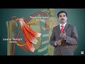 AXILLARY ARTERY ANATOMY ANIMATED LECTURE
 Abdominal aorta - Origin , course , branches - USMLE Step 1 Videos
 Liver Anatomy - Accessory Organs Part 1
 Anatomy of Stomach ( Surgery ) : Embryology , Gross anatomy , Histology , Arterial and Nerve supply
 The Sciatic Nerve Anatomy - Origin, Course, Relations, Branches, Distribution and Clinical anatomy
 Appendix Problem Cause Sign Symptom Treatment Medicine Surgery | Best Laparoscopic Key Hole Surgery
 Abdominal organs (plastic anatomy)
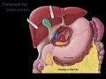 Peritoneum tutorial
 Vermiform Appendix ( Features & Positions )
 LARGE INTESTINE | ANATOMY | SIMPLIFIED
 Anatomy of the VERMIFORM APPENDIX || Dr. Yusuf ||
 Anatomy of Large Intestine (Structures and Walls)