Reporting a lumbar spine MRI - disc extrusion
Lumbar spine MRI is probably one of the most commonly performed MRI exams in radiology. This is because back pain is extraordinarily common and increases with age. As a result, we do a number of studies to try to determine what is the cause of the back pain.
There are a number of things that can cause back pain. Discs can bulge (and protrude, or extrude), facets can degenerate, and endplates can erode. All of these can cause pain. Unfortunately, the nerves of the spinal cord and extremities pass through this region and their compression can cause a lot of pain.
Today we'll do things a little differently. I'm going to show you an example case on the radiology PACS of a young patient with back pain radiating down the right leg. I'll show you how I set up the study to view and systematically how I look at it. In addition, I'll show you how our reports are formatted and how you can as well.
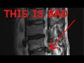
This patient has a disc extrusion at L4-L5 which is pressing on the exiting nerve root in the lateral recess (or subarticular zone). This is just one of the many reasons to have radicular pain.
The level of this video is appropriate for medical students, junior residents, and trainees in other specialties who have an interest in spine imaging. General medicine practitioners will see plenty of back pain and may wonder what we are thinking. Neurosurgeons and orthopedic surgeons can learn more to strengthen their practice as well.
Check out this video and additional content on http://www.learnneuroradiology.com
Видео Reporting a lumbar spine MRI - disc extrusion канала LearnNeuroradiology
There are a number of things that can cause back pain. Discs can bulge (and protrude, or extrude), facets can degenerate, and endplates can erode. All of these can cause pain. Unfortunately, the nerves of the spinal cord and extremities pass through this region and their compression can cause a lot of pain.
Today we'll do things a little differently. I'm going to show you an example case on the radiology PACS of a young patient with back pain radiating down the right leg. I'll show you how I set up the study to view and systematically how I look at it. In addition, I'll show you how our reports are formatted and how you can as well.
This patient has a disc extrusion at L4-L5 which is pressing on the exiting nerve root in the lateral recess (or subarticular zone). This is just one of the many reasons to have radicular pain.
The level of this video is appropriate for medical students, junior residents, and trainees in other specialties who have an interest in spine imaging. General medicine practitioners will see plenty of back pain and may wonder what we are thinking. Neurosurgeons and orthopedic surgeons can learn more to strengthen their practice as well.
Check out this video and additional content on http://www.learnneuroradiology.com
Видео Reporting a lumbar spine MRI - disc extrusion канала LearnNeuroradiology
Показать
Комментарии отсутствуют
Информация о видео
Другие видео канала
 How to Read a Spine MRI
 Head and neck anatomy landmarks
 Artrosis lumbar - Signos radiológicos
 Chest Radiography: Nodules, Masses, and Lung Cancer
 Low Back Pain , Disc Herniation - Everything You Need To Know - Dr. Nabil Ebraheim
 Lumbar Spine MRI by Eric Tranvinh, MD, Stanford Radiology
 Can a Disc Herniation Heal Itself?
 Vascular Imaging of the Head and Neck - Case C
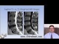 Dr. Gillard lectures on How to Read Your Lumbar MRI
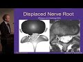 Lumbar MRI: What the Findings Mean and How They Should Be Reported - J. Jarvik, MD, MPH
 Multiple sclerosis – white spots and red flags - part 2 - Mimics and Variants
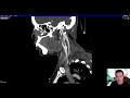 Vascular Imaging of the Head and Neck - Case B
 How to identify a vertebra (anatomy)
 Which Meningiomas Should Not Be Treated by Dr. Steven D. Chang
 Introduction to Radiology: Magnetic Resonance Imaging
 Dr. Gillard Lectures on Lumbar Disc Herniation / Protrusion. Part I of II
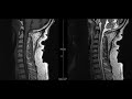 Noncontrast MRI cervical spine search pattern
 Treatment for lumbar spine disc bulge and sciatica - wk 1 | Feat. Tim Keeley | No.58 | Physio REHAB
 10 Exercises to Avoid With Sciatica (Bulging or Herniated Disc) or Back Pain.
 Lumbar Disc Herniation MRI Explained | Dr. Jeffrey P. Johnson | HD