Gel Electrophoresis of DNA
#rethinkbiology #dnagelelectrophoresis #gelelectrophoresisanimation
Gel electrophoresis
If you break the word into electro and phoresis, electro refers to the energy of electricity and phoresis mean to carry across. So, the electrophoresis is a method for separation of charged macromolecules like DNA RNA proteins and their fragments based on their size. The gel is used as a medium for the macromolecules to move, that's why it is called gel electrophoresis.
Gel electrophoresis of DNA
The basic principle of gel electrophoresis is running a current through the gel loaded with the macromolecule of our interest. Based on their size the molecule will travel through the gel at different speeds so this will allow them to be separated from one another. It can be based on the charge also if there is a difference in charge among them. But in case of DNA, all DNA molecules have the same charge to mass ratio and they are negatively charged. Separation also depends on the compactness of the DNA, the more compact DNA will able to migrate first through the gel. The gel is a jelly-like material, which is composed of tiny molecules and they held together by hydrogen bonds to form a sieve-like structure with pores. Within the gel, the larger and less compact fractions of DNA move slowly while the smaller and compact pieces move quickly through the gel.
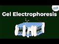
Two types of gels are commonly used in separation of DNA. Polyacrylamide and agarose gel. At one end of the gel, some wells are made to load the DNA samples. The gel is first placed in a box, one end of the box is hooked to a positive electrode and the other end to a negative electrode. The box is then filled with a salt containing buffer solution which can conduct electricity. The end of the gel with wells will be placed near the negative electrode. As DNA is negatively charged it will move towards the positive electrode. To track the DNA movement a tracking dye is usually mixed with that samples. DNA is nearly transparent it can’t be observed inside the gel. Bromophenol blue and xylene cyanol are used as tracking dyes.
The tracking dye will always run a little ahead of the smallest fragment of DNA. Also, a DNA staining dye is generally mixed with the gel to visualize the separated DNA fragments after the run ends. To know the size of the different DNA fragments in the samples we need to run a DNA ladder in one of the wells. DNA ladder is just like a DNA sample but its fragment size is defined. So after the run completes the sizes of the DNA fragments in the test samples can easily be calculated comparing it with the DNA ladder. After completion of the run, the gel can be taken out and DNA can be visualised by placing it under UV light.
Gel electrophoresis of the DNA can be used to estimate the size of DNA fragments following restriction enzyme digestion. It can also be done to analyse the PCR products. It can be done to check for a match between the DNA sample collected from the crime spot and the DNA of suspected individuals.
Видео Gel Electrophoresis of DNA канала Rethink Biology
Gel electrophoresis
If you break the word into electro and phoresis, electro refers to the energy of electricity and phoresis mean to carry across. So, the electrophoresis is a method for separation of charged macromolecules like DNA RNA proteins and their fragments based on their size. The gel is used as a medium for the macromolecules to move, that's why it is called gel electrophoresis.
Gel electrophoresis of DNA
The basic principle of gel electrophoresis is running a current through the gel loaded with the macromolecule of our interest. Based on their size the molecule will travel through the gel at different speeds so this will allow them to be separated from one another. It can be based on the charge also if there is a difference in charge among them. But in case of DNA, all DNA molecules have the same charge to mass ratio and they are negatively charged. Separation also depends on the compactness of the DNA, the more compact DNA will able to migrate first through the gel. The gel is a jelly-like material, which is composed of tiny molecules and they held together by hydrogen bonds to form a sieve-like structure with pores. Within the gel, the larger and less compact fractions of DNA move slowly while the smaller and compact pieces move quickly through the gel.
Two types of gels are commonly used in separation of DNA. Polyacrylamide and agarose gel. At one end of the gel, some wells are made to load the DNA samples. The gel is first placed in a box, one end of the box is hooked to a positive electrode and the other end to a negative electrode. The box is then filled with a salt containing buffer solution which can conduct electricity. The end of the gel with wells will be placed near the negative electrode. As DNA is negatively charged it will move towards the positive electrode. To track the DNA movement a tracking dye is usually mixed with that samples. DNA is nearly transparent it can’t be observed inside the gel. Bromophenol blue and xylene cyanol are used as tracking dyes.
The tracking dye will always run a little ahead of the smallest fragment of DNA. Also, a DNA staining dye is generally mixed with the gel to visualize the separated DNA fragments after the run ends. To know the size of the different DNA fragments in the samples we need to run a DNA ladder in one of the wells. DNA ladder is just like a DNA sample but its fragment size is defined. So after the run completes the sizes of the DNA fragments in the test samples can easily be calculated comparing it with the DNA ladder. After completion of the run, the gel can be taken out and DNA can be visualised by placing it under UV light.
Gel electrophoresis of the DNA can be used to estimate the size of DNA fragments following restriction enzyme digestion. It can also be done to analyse the PCR products. It can be done to check for a match between the DNA sample collected from the crime spot and the DNA of suspected individuals.
Видео Gel Electrophoresis of DNA канала Rethink Biology
Показать
Комментарии отсутствуют
Информация о видео
Другие видео канала
 What is Gel Electrophoresis | Don't Memorise
 DNA Replication Made Easy
 DNA Sequencing - 3D
 SDS - PAGE
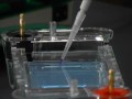 Principles of Gel Electrophoresis
 Gel Electrophoresis
 The Principle of Agarose Gel Electrophoresis, a full explanatory video
 Drew Berry: Animations of unseeable biology
 DNA extraction from Blood
 Chaos: The Science of the Butterfly Effect
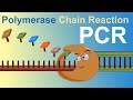 PCR - Polymerase Chain Reaction (IQOG-CSIC)
 Gel Electrophoresis
 Southern Blotting
 Holliday Model of Recombination
 SDS PAGE | polyacrylamide gel electrophoresis
 Southern blotting
 DNA Fingerprinting | Genetics | Biology | FuseSchool
 Agarose Gel Electrophoresis to separate DNA fragments
 DNA gel electrophoresis lab demo
 DNA Replication (Updated)