Pivot Shift Test - Everything You Need To Know - Dr. Nabil Ebraheim
Dr. Ebraheim’s educational animated video demonstrates the Pivot Shift test.
The anterior cruciate ligament is located in the front of the knee. Rupture of the ACL is a condition commonly seen in sports usually due to a non-contact pivoting injury. The Pivot shift test is a specific test for ACL deficient knee (ACL injury). Pivot shift is pathognomonic for an ACL tear and is best demonstrated in a chronic setting. Lachman’s test is the most sensitive test for ACL injury. The ACL keeps the tibia from sliding pout in front of the femur and provides rotational stability to the knee. The tibia moves anterolaterally in extension, however when you flex the knee the IT band becomes a flexor of the knee. The IT band pulls back and reduces the tibia. Rupture of the ACL causes anterolateral rotatory instability. The Pivot shift test goes from extension (tibia subluxed) to flexion, with the tibia reduced by the iliotibial band.
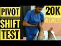
How do you perform the Pivot shift test? Both the Lachman’s test and the Pivot shift test are associated with 20-30 degrees of knee flexion. The Lachman’s test starts at 20-30 degrees of flexion. With the Pivot shift test you feel the clunk at 20-30 degrees of flexion. 20-30 degrees of flexion is important for examination of the ACL (remember that).
The femur is stabilized with one hand and the other hand pulls the tibia anteriorly and posteriorly against the femur. The tibia can be pulled forward more than normal (anterior translation). The examiner will have a sense of increased movement and lack of a solid end point. The patient should be lying supine. Make sure the patient is totally relaxed. With Pivot shift, the knee is in the subluxed position when the knee is in full extension. The Pivot shift starts with extension of the knee and you can feel a clunk at 20-30 degrees of flexion. Hold the knee in full extension then add valgus force plus internal rotation of the tibia to increase the rotational instability of the knee. Then take the knee into flexion. The iliotibial band will reduce the tibia and create the clunk on the outside of the knee. Always compare with the other side. The ACL prevents anterior translation of the tibia. It is secondary restraint to tibial rotation and varus and valgus.
The ACL consists of two bundles: Posterolateral bundle: prevents pivot shift, contributes to rotational stability, and also prevents internal rotation of the tibia with the knee in near extension (tight in extension, loose in flexion). If it is sectioned, it increases the anterior translation and tibial rotation at 30 degrees of flexion.
Anteromedial bundle: tight in flexion, if it is sectioned, it increases anterior translation at 90 degrees of flexion.
In pivot shift, the knee subluxes in extension, and reduces at 20-30 degrees of flexion. The Pivot shift correlated closely with patient satisfaction of their reconstructed knee. It is a measure of functional instability following ACL reconstruction.
Vertical femoral tunnel placement will cause a rotational instability seen as positive pivot shift and the malposition of the bone tunnel will be seen in an AP view x-ray of the knee. The 9 or 10 o’clock position is better than the 12 o’clock. Vertical position is bad.
The patient with an ACL injury usually has a non-contact pivoting injury event with:
•Awkward landing
•Feeling a “pop” sensation
•Immediate swelling
•Aspiration usually shows blood in the knee (75% chance of ACL tear with hemorrhage in the knee)
•Positive Lachman’s test (may be hard to examine because of the pain).
Radiological exam
Get an MRI. MRI of the knee joint shows bone lesions or bruising in the typical location associated with tears of the ACL. These injuries are typically located at the middle of the femoral condyle and posterior part of the tibia laterally. You may find a triple injury within the MRI (O’Donoghue’s unhappy triad): medial collateral ligament injury, anterior cruciate ligament injury, and meniscus injury.
In chronic ACL tears, the posterior horn of the medial meniscus is the most commonly injured structure.
In acute ACL tear, send the patient for therapy for range of motion, brace the patient and allow the MCL to heal and reconstruct the ACL later if needed. Stress hamstring therapy in ACL tear. The patient will probably complain of instability immediately or later.
Become a friend on facebook:
http://www.facebook.com/drebraheim
Follow me on twitter:
https://twitter.com/#!/DrEbraheim_UTMC
Donate to the University of Toledo Foundation Department of Orthopaedic Surgery Endowed Chair Fund:
https://www.utfoundation.org/foundation/home/Give_Online.aspx?sig=29
Background music provided as a free download from YouTube Audio Library.
Song Title: Every Step
Видео Pivot Shift Test - Everything You Need To Know - Dr. Nabil Ebraheim канала nabil ebraheim
The anterior cruciate ligament is located in the front of the knee. Rupture of the ACL is a condition commonly seen in sports usually due to a non-contact pivoting injury. The Pivot shift test is a specific test for ACL deficient knee (ACL injury). Pivot shift is pathognomonic for an ACL tear and is best demonstrated in a chronic setting. Lachman’s test is the most sensitive test for ACL injury. The ACL keeps the tibia from sliding pout in front of the femur and provides rotational stability to the knee. The tibia moves anterolaterally in extension, however when you flex the knee the IT band becomes a flexor of the knee. The IT band pulls back and reduces the tibia. Rupture of the ACL causes anterolateral rotatory instability. The Pivot shift test goes from extension (tibia subluxed) to flexion, with the tibia reduced by the iliotibial band.
How do you perform the Pivot shift test? Both the Lachman’s test and the Pivot shift test are associated with 20-30 degrees of knee flexion. The Lachman’s test starts at 20-30 degrees of flexion. With the Pivot shift test you feel the clunk at 20-30 degrees of flexion. 20-30 degrees of flexion is important for examination of the ACL (remember that).
The femur is stabilized with one hand and the other hand pulls the tibia anteriorly and posteriorly against the femur. The tibia can be pulled forward more than normal (anterior translation). The examiner will have a sense of increased movement and lack of a solid end point. The patient should be lying supine. Make sure the patient is totally relaxed. With Pivot shift, the knee is in the subluxed position when the knee is in full extension. The Pivot shift starts with extension of the knee and you can feel a clunk at 20-30 degrees of flexion. Hold the knee in full extension then add valgus force plus internal rotation of the tibia to increase the rotational instability of the knee. Then take the knee into flexion. The iliotibial band will reduce the tibia and create the clunk on the outside of the knee. Always compare with the other side. The ACL prevents anterior translation of the tibia. It is secondary restraint to tibial rotation and varus and valgus.
The ACL consists of two bundles: Posterolateral bundle: prevents pivot shift, contributes to rotational stability, and also prevents internal rotation of the tibia with the knee in near extension (tight in extension, loose in flexion). If it is sectioned, it increases the anterior translation and tibial rotation at 30 degrees of flexion.
Anteromedial bundle: tight in flexion, if it is sectioned, it increases anterior translation at 90 degrees of flexion.
In pivot shift, the knee subluxes in extension, and reduces at 20-30 degrees of flexion. The Pivot shift correlated closely with patient satisfaction of their reconstructed knee. It is a measure of functional instability following ACL reconstruction.
Vertical femoral tunnel placement will cause a rotational instability seen as positive pivot shift and the malposition of the bone tunnel will be seen in an AP view x-ray of the knee. The 9 or 10 o’clock position is better than the 12 o’clock. Vertical position is bad.
The patient with an ACL injury usually has a non-contact pivoting injury event with:
•Awkward landing
•Feeling a “pop” sensation
•Immediate swelling
•Aspiration usually shows blood in the knee (75% chance of ACL tear with hemorrhage in the knee)
•Positive Lachman’s test (may be hard to examine because of the pain).
Radiological exam
Get an MRI. MRI of the knee joint shows bone lesions or bruising in the typical location associated with tears of the ACL. These injuries are typically located at the middle of the femoral condyle and posterior part of the tibia laterally. You may find a triple injury within the MRI (O’Donoghue’s unhappy triad): medial collateral ligament injury, anterior cruciate ligament injury, and meniscus injury.
In chronic ACL tears, the posterior horn of the medial meniscus is the most commonly injured structure.
In acute ACL tear, send the patient for therapy for range of motion, brace the patient and allow the MCL to heal and reconstruct the ACL later if needed. Stress hamstring therapy in ACL tear. The patient will probably complain of instability immediately or later.
Become a friend on facebook:
http://www.facebook.com/drebraheim
Follow me on twitter:
https://twitter.com/#!/DrEbraheim_UTMC
Donate to the University of Toledo Foundation Department of Orthopaedic Surgery Endowed Chair Fund:
https://www.utfoundation.org/foundation/home/Give_Online.aspx?sig=29
Background music provided as a free download from YouTube Audio Library.
Song Title: Every Step
Видео Pivot Shift Test - Everything You Need To Know - Dr. Nabil Ebraheim канала nabil ebraheim
Показать
Комментарии отсутствуют
Информация о видео
Другие видео канала
 Pivot Shift Test for ACL
 Heel Pain- Everything You Need To Know - Dr. Nabil Ebraheim
 McMurray's Test - Everything You Need To Know - Dr. Nabil Ebraheim
 Valgus Stress Test | Medial Collateral Ligament (MCL) Injury
 The Lateral Pivot-Shift Test for Anterior Cruciate Ligament Rupture
 ACL Tears Radiological Evaluation - Everything You Need To Know - Dr. Nabil Ebraheim
 Reverse Pivot-Shift Test | Posterolateral Rotatory Instability of the Knee
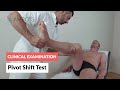 Pivot Shift Test - Clinical Examination
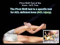 Pivot Shift Test ACL Tear - Everything You Need To Know - Dr. Nabil Ebraheim
 Meniscal Tears Examination & Tests - Everything You Need To Know - Dr. Nabil Ebraheim
 Pivot Shift Test | Anterior Cruciate Ligament (ACL) Rupture Knee
 Ankle Ligament Injury, evaluation and tests - Everything You Need To Know - Dr. Nabil Ebraheim
 Apley's Test for Knee | Clinical Physio Premium
 Tests For Examination Of The Knee - Everything You Need To Know - Dr. Nabil Ebraheim
 Test de Macmurray
 Posterior Sag Sign | Posterior Cruciate Ligament Tear
 Knee Joint Full Assessment Run Through | Clinical Physio
 ACL Evaluation:Positive Lachman's Test & Positive Reverse Pivot Shift Test
 Pivot Shift Test - YouTube
 Quadriceps Active Test / Active Drawer Test | Posterior Cruciate Ligament Tear