VERTEBRAL COLUMN ANATOMY (2/2) - Ligaments and the Spinal Cord
Three major ligaments of the spine that allow flexion and extension of the spine while keeping the bones aligned – the ligamentum flavum, anterior longitudinal ligament, and the posterior longitudinal ligament. The anterior and posterior longitudinal ligaments are continuous bands that run from the top to the bottom of the vertebral column and prevent excess movement. The ligament flavum attaches between lamina of each vertebra.
Some additional ligaments I’d like to point out are the intertransverse ligament, the supraspinous ligament, and the interspinous ligament. The intertransverse ligaments stretch between the transverse processes of the spine. The supraspinous ligament is found along the vertebral column, connecting the tips of the spinous processes from the cervical vertebra to the sacrum. At the seventh vertebra, the supraspinous ligament is continuous with the nuchal ligament, which runs from the seventh vertebra to the external occipital protuberance of the skull. The interspinous ligaments are thin, membranous ligaments stretching between adjacent spinous processes.
Finally, I’d like to briefly discuss the spinal cord, which is floating in cerebrospinal fluid within the dural tube, which is inside the vertebral arch. The spinal cord is about the thickness of your thumb. At around 18 inches long, it runs from the brainstem to the 1st or 2nd lumbar vertebra within the spinal canal, ending in the conus medullaris. Extending from the conus medullaris is the cauda equina – Latin for horse tail because it is a bunch of spinal nerves that very much looks like a tail. The cauda equina occupies the lumbar cistern – a space beneath the conus medullaris. The filum terminale extends from the end of the spinal chord and anchors it to the tailbone.
31 pairs of spinal nerves branch off from the spinal cord. Each spinal nerve has 2 roots – one ventral for motor impulses from brain, and one dorsal for sensory impulses to brain. Ventral and dorsal roots fuse to form spinal nerve, which exits the vertebral column via the intervertebral foramen between the vertebrae.
3D model from:
https://www.turbosquid.com/3d-models/human-torso-muscles-max/647193
Видео VERTEBRAL COLUMN ANATOMY (2/2) - Ligaments and the Spinal Cord канала Neural Academy
Показать
Комментарии отсутствуют
Информация о видео
Другие видео канала
 Vertebral Column – Anatomy | Lecturio
 Posterior Lumbar Interbody Fusion Overview
 Individual Vertebrae with Structures
 Neurology - Spinal Cord Introduction
 Spine anatomy - learn from a NEUROSURGEON!
 Lumbar Spine Anatomy
 The Major Ligaments of the Spine
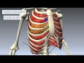 Muscles of the Thoracic Wall - 3D Anatomy Tutorial
 Spinal Trauma: Cervical Trauma Protocol, Common Spinal Fractures – Radiology | Lecturio
 Skull bones, sutures and landmarks
 Erector spinae (back muscles)
 Spinal Cord - External Anatomy - 3D Anatomy Tutorial
 Ligaments of Vertebral Column Anatomy (Nuchal, Interspinous, Supraspinous)
 Vertebral ligaments
 Intervertebral disc (anatomy)
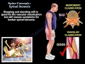 Lumbar Spinal Stenosis - Everything You Need To Know - Dr. Nabil Ebraheim
 VERTEBRAL COLUMN ANATOMY (1/2)
 Thoracic vertebrae vs. Lumbar vertebrae - Human Anatomy | Kenhub
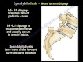 Spondylolysis,Spondylolisthesis,Spondylitis&Spondylosis-EverythingYou Need To Know-Dr.Nabil Ebraheim
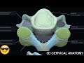 Cervical Vertebrae Anatomy || 3D #OMT #COMLEX