- Популярные видео
- Авто
- Видео-блоги
- ДТП, аварии
- Для маленьких
- Еда, напитки
- Животные
- Закон и право
- Знаменитости
- Игры
- Искусство
- Комедии
- Красота, мода
- Кулинария, рецепты
- Люди
- Мото
- Музыка
- Мультфильмы
- Наука, технологии
- Новости
- Образование
- Политика
- Праздники
- Приколы
- Природа
- Происшествия
- Путешествия
- Развлечения
- Ржач
- Семья
- Сериалы
- Спорт
- Стиль жизни
- ТВ передачи
- Танцы
- Технологии
- Товары
- Ужасы
- Фильмы
- Шоу-бизнес
- Юмор
Lateral Unicondylar Knee Arthroplasty
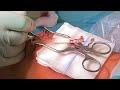
This video demonstrates the surgical technique for lateral unicompartmental knee arthroplasty.
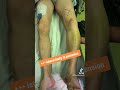
The procedure is performed under general or epidural anesthesia, with the patient positioned on a standard operating table using two leg holders. The knee is flexed to 90 degrees.
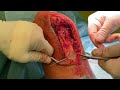
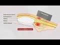
The skin incision starts at the superior pole of the patella and extends distally toward the lateral aspect of the tibial tuberosity, ending approximately two centimeters below the joint line.
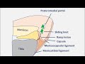
A lateral arthrotomy is performed, and the joint is opened. The lateral portion of the fat pad is excised to expose the lateral femoral condyle, tibial plateau, and anterior cruciate ligament. The ACL integrity, as well as the medial and patellofemoral compartments, are carefully assessed. Osteophytes in the intercondylar notch are removed to prevent impingement, while lateral femoral osteophytes are preserved as landmarks.
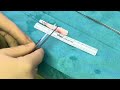
The tibial cut is then performed. Resection should be minimal, typically 2 to 4 millimeters, as the disease predominantly affects the femoral side. An extramedullary guide is used, and the natural tibial slope, close to zero degrees, should be reproduced. The sagittal cut follows a line respecting the tibial spine eminence, with careful retraction of the patellar tendon.
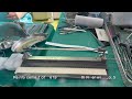
Femoral preparation follows. The distal cut is made using a cutting guide connected to a spacer.
For the posterior and oblique cut and the peg holes a specific cutting guide is used.
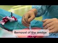
Rotation of this cutting guide is critical and should follow the lateral condylar anatomy to avoid internal malrotation in extension. Ther should be a distance of 1-2 mm to the anterior cartilage. After peg hole drilling, posterior snd oblique cut, posterior osteophytes are removed to ensure full flexion.
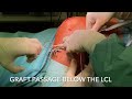
The tibial component is sized to achieve optimal coverage without overhang. Trial components are inserted, and flexion and extension gaps are assessed. Alignment and potential impingement must be carefully checked.
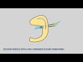
Final implantation is performed with cementation of the tibial component first, followed by inserting the polyethylene insert and the femoral component.
Understanding the screw-home mechanism is essential. External tibial rotation during terminal extension contributes to stability, and femoral positioning must account for this to avoid impingement.
This technique emphasizes precise alignment, conservative resection, and restoration of native knee biomechanics.
#knee #orthopedics #arthroscopy #kneesurgery #kneepain #kneesurgey #cartilage #patella #medicine #kneearthroplasty #partialkneereplacement #lateralunicondylarkneearthroplasty
Видео Lateral Unicondylar Knee Arthroplasty канала Prof. Wolf Petersen
The procedure is performed under general or epidural anesthesia, with the patient positioned on a standard operating table using two leg holders. The knee is flexed to 90 degrees.
The skin incision starts at the superior pole of the patella and extends distally toward the lateral aspect of the tibial tuberosity, ending approximately two centimeters below the joint line.
A lateral arthrotomy is performed, and the joint is opened. The lateral portion of the fat pad is excised to expose the lateral femoral condyle, tibial plateau, and anterior cruciate ligament. The ACL integrity, as well as the medial and patellofemoral compartments, are carefully assessed. Osteophytes in the intercondylar notch are removed to prevent impingement, while lateral femoral osteophytes are preserved as landmarks.
The tibial cut is then performed. Resection should be minimal, typically 2 to 4 millimeters, as the disease predominantly affects the femoral side. An extramedullary guide is used, and the natural tibial slope, close to zero degrees, should be reproduced. The sagittal cut follows a line respecting the tibial spine eminence, with careful retraction of the patellar tendon.
Femoral preparation follows. The distal cut is made using a cutting guide connected to a spacer.
For the posterior and oblique cut and the peg holes a specific cutting guide is used.
Rotation of this cutting guide is critical and should follow the lateral condylar anatomy to avoid internal malrotation in extension. Ther should be a distance of 1-2 mm to the anterior cartilage. After peg hole drilling, posterior snd oblique cut, posterior osteophytes are removed to ensure full flexion.
The tibial component is sized to achieve optimal coverage without overhang. Trial components are inserted, and flexion and extension gaps are assessed. Alignment and potential impingement must be carefully checked.
Final implantation is performed with cementation of the tibial component first, followed by inserting the polyethylene insert and the femoral component.
Understanding the screw-home mechanism is essential. External tibial rotation during terminal extension contributes to stability, and femoral positioning must account for this to avoid impingement.
This technique emphasizes precise alignment, conservative resection, and restoration of native knee biomechanics.
#knee #orthopedics #arthroscopy #kneesurgery #kneepain #kneesurgey #cartilage #patella #medicine #kneearthroplasty #partialkneereplacement #lateralunicondylarkneearthroplasty
Видео Lateral Unicondylar Knee Arthroplasty канала Prof. Wolf Petersen
Комментарии отсутствуют
Информация о видео
5 апреля 2026 г. 12:58:51
00:04:04
Другие видео канала