- Популярные видео
- Авто
- Видео-блоги
- ДТП, аварии
- Для маленьких
- Еда, напитки
- Животные
- Закон и право
- Знаменитости
- Игры
- Искусство
- Комедии
- Красота, мода
- Кулинария, рецепты
- Люди
- Мото
- Музыка
- Мультфильмы
- Наука, технологии
- Новости
- Образование
- Политика
- Праздники
- Приколы
- Природа
- Происшествия
- Путешествия
- Развлечения
- Ржач
- Семья
- Сериалы
- Спорт
- Стиль жизни
- ТВ передачи
- Танцы
- Технологии
- Товары
- Ужасы
- Фильмы
- Шоу-бизнес
- Юмор
Double Level Osteotomy In Three Minutes
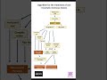
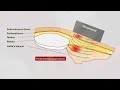
Double-level osteotomy (DLO) is indicated in patients with varus malalignment originating from both the distal femur and proximal tibia, particularly when isolated tibial correction would result in excessive joint line obliquity. Preoperative planning is performed using long-leg weight-bearing radiographs to determine the mechanical axis deviation and to quantify the femoral and tibial components of the deformity. Correction is distributed between the distal femur and proximal tibia to restore physiological alignment while maintaining a horizontal joint line orientation.
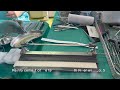
The procedure is performed with the patient in the supine position under general or regional anesthesia. Fluoroscopy is used throughout the procedure.
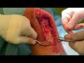
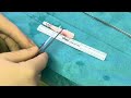
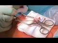
The distal femoral osteotomy is performed first. Through a lateral approach, the iliotibial band is incised and the vastus lateralis is elevated anteriorly to expose the lateral distal femur. Under fluoroscopic guidance, a guide wire is inserted toward the medial cortex at the level of the planned hinge. This wire serves as a hinge protection wire and helps prevent fracture of the medial cortical hinge during closure of the osteotomy. Two additional guide wires are then inserted to define the osteotomy plane and the planned wedge resection. A lateral closing-wedge osteotomy is performed using an oscillating saw while preserving the medial cortical hinge. The bone wedge is removed and the osteotomy is gradually closed under fluoroscopic control until the planned correction is achieved. Fixation is performed using a precontoured lateral distal femoral locking plate.
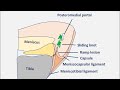
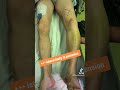
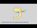
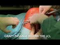
Subsequently, a medial opening-wedge high tibial osteotomy is performed. Through an anteromedial approach to the proximal tibia, the superficial medial collateral ligament is carefully released from the tibial cortex. A Hohmann retractor is placed posteriorly to protect the neurovascular structures. Under fluoroscopic guidance, two guide wires are inserted toward the lateral hinge, which is located just above the fibular head. A biplanar osteotomy is then created using an oscillating saw while leaving approximately 5–10 mm of the lateral cortex intact. The osteotomy gap is gradually opened using chisels or a spreading device until the planned correction is reached. Limb alignment is checked intraoperatively using a long alignment rod or fluoroscopic assessment of the mechanical axis.
The tibial osteotomy is stabilized with an angular stable plate. Bone graft or bone substitute may be used depending on the size of the opening wedge.
Finally, overall limb alignment and joint line orientation are verified fluoroscopically to confirm restoration of physiological mechanical axis alignment. Postoperatively, patients are mobilized with partial weight-bearing for approximately six weeks. Immediate range-of-motion exercises are initiated, followed by gradual progression to full weight-bearing after radiographic signs of osteotomy healing.
#knee #orthopedics #kneesurgey #medicine #arthroscopy #kneepain #kneesurgery #patella #osteotomy #osteoarthritis
Видео Double Level Osteotomy In Three Minutes канала Prof. Wolf Petersen
The procedure is performed with the patient in the supine position under general or regional anesthesia. Fluoroscopy is used throughout the procedure.
The distal femoral osteotomy is performed first. Through a lateral approach, the iliotibial band is incised and the vastus lateralis is elevated anteriorly to expose the lateral distal femur. Under fluoroscopic guidance, a guide wire is inserted toward the medial cortex at the level of the planned hinge. This wire serves as a hinge protection wire and helps prevent fracture of the medial cortical hinge during closure of the osteotomy. Two additional guide wires are then inserted to define the osteotomy plane and the planned wedge resection. A lateral closing-wedge osteotomy is performed using an oscillating saw while preserving the medial cortical hinge. The bone wedge is removed and the osteotomy is gradually closed under fluoroscopic control until the planned correction is achieved. Fixation is performed using a precontoured lateral distal femoral locking plate.
Subsequently, a medial opening-wedge high tibial osteotomy is performed. Through an anteromedial approach to the proximal tibia, the superficial medial collateral ligament is carefully released from the tibial cortex. A Hohmann retractor is placed posteriorly to protect the neurovascular structures. Under fluoroscopic guidance, two guide wires are inserted toward the lateral hinge, which is located just above the fibular head. A biplanar osteotomy is then created using an oscillating saw while leaving approximately 5–10 mm of the lateral cortex intact. The osteotomy gap is gradually opened using chisels or a spreading device until the planned correction is reached. Limb alignment is checked intraoperatively using a long alignment rod or fluoroscopic assessment of the mechanical axis.
The tibial osteotomy is stabilized with an angular stable plate. Bone graft or bone substitute may be used depending on the size of the opening wedge.
Finally, overall limb alignment and joint line orientation are verified fluoroscopically to confirm restoration of physiological mechanical axis alignment. Postoperatively, patients are mobilized with partial weight-bearing for approximately six weeks. Immediate range-of-motion exercises are initiated, followed by gradual progression to full weight-bearing after radiographic signs of osteotomy healing.
#knee #orthopedics #kneesurgey #medicine #arthroscopy #kneepain #kneesurgery #patella #osteotomy #osteoarthritis
Видео Double Level Osteotomy In Three Minutes канала Prof. Wolf Petersen
Комментарии отсутствуют
Информация о видео
7 марта 2026 г. 23:41:14
00:03:01
Другие видео канала