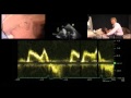
Echocardiography Essentials: Evaluating right ventricular size and function
After watching this video, you will be able to recognize right ventricular dilatation, significant hypertrophy and hyper- and hypokinesis using transthoracic echo. You will also know how to tell if pulmonary embolism is suspected.
This video was taken from our hands-on and CME accredited Echocardiography Essentials Course. You can check it out here:
https://www.medmastery.com/course/echocardiography-essentials
Видео Echocardiography Essentials: Evaluating right ventricular size and function канала Medmastery
This video was taken from our hands-on and CME accredited Echocardiography Essentials Course. You can check it out here:
https://www.medmastery.com/course/echocardiography-essentials
Видео Echocardiography Essentials: Evaluating right ventricular size and function канала Medmastery
Показать
Комментарии отсутствуют
Информация о видео
Другие видео канала
 Diastolic Function — A Simple Approach
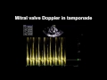 Echocardiography Essentials: Detecting pericardial effusions
 Right Ventricular Assessment: Move Over RV S' & TAPSE, Strain all the Way – Prof Liza Thomas
 Echocardiography Essentials: Detecting aortic regurgitation
 Assessment of Right Ventricle (THOMAS DI SALVO, MD) May 17, 2018
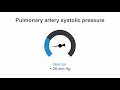 How to estimate pulmonary artery systolic pressure (PASP) using echo
 Hypertrophic Obstructive Cardiomyopathy (HOCM)
 Echocardiography Essentials: Spotting tricuspid and pulmonary valve disease
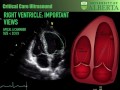 Right Ventricular Function in Critical Illness
 Pitfalls of VTI
 Echo BachelorClass - Your introduction to basic echocardiography
 Point of Care Echo: Stroke Volume Determination
 Assessing Right Ventricular Function - ECHO Course l The EKG Guy - www.ekg.md
 Evaluation and Management of Mitral Regurgitation | 1: Guidelines and Quantification
 Point-of-Care Echo: Regional Wall Motion Abnormalities
 Echocardiographic Assesment of Mitral Regurgitation
 Joint Echo Conference: Mitral Stenosis
 Estimate the RVSP (Right Ventricular Systolic Pressure) or PASP. Perioperative & Critical Care POCUS
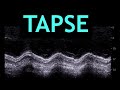 All about TAPSE! (Echocardiography)
 Tissue Doppler Step by Step - Medial e' Example