Tissue Doppler Step by Step - Medial e' Example
Learn how to use Tissue Wave Doppler in a step by step fashion. In this example, we will be using Tissue Doppler imaging to measure the medial annulus e' (this is used to access diastolic dysfunction).
These same steps can be used to apply Tissue Doppler to any other applications you may feel necessary.
Visit: https://pocus101.com/knobology for a complete ultrasound Knobology and Machine settings tutorial we made for you!
Видео Tissue Doppler Step by Step - Medial e' Example канала POCUS 101
These same steps can be used to apply Tissue Doppler to any other applications you may feel necessary.
Visit: https://pocus101.com/knobology for a complete ultrasound Knobology and Machine settings tutorial we made for you!
Видео Tissue Doppler Step by Step - Medial e' Example канала POCUS 101
Показать
Комментарии отсутствуют
Информация о видео
Другие видео канала
 Tissue Doppler Imaging in Low Risk Chest Pain Patients
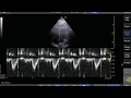 Pulse Wave Doppler Step by Step - LVOT VTI Example
 Basic Transthoracic Echocardiography (Cardiac Ultrasound) - Made Simple
 Diastolic Function — A Simple Approach
 Tissue Doppler LV
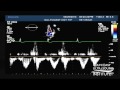 Hot Tips - Calculating the Aortic Valve Area Using the Continuity Equation
 Doppler Ultrasound Part 1 - Principles (w/ focus on Spectral Waveforms)
 How to estimate pulmonary artery systolic pressure (PASP) using echo
 Transthoracic Echo full protocol. Part II: Parasternal View (PLAX , PSAX, RVIT, RVOT, M-Mode)
 How to use Color Doppler on Ultrasound - Step by Step Guide
 E/A Ratio and Diastolic Dysfunction
 Estimating Ejection Fraction with Point of Care Echo
 Echo MasterClass - Your introduction to advanced echocardiography
 21. Tissue doppler imaging of the free wall of the RV at the tricuspid annulus
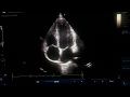 MAPSE and TAPSE
 Echocardiography of MR
 Basic TTE Video Tutorial
 Ultrasound Podcast - Tissue Harmonics
 Introduction to CT Head: Approach and Principles
 Echocardiography Essentials: Evaluating right ventricular size and function