Nerve Regeneration - Everything You Need To Know - Dr. Nabil Ebraheim
Educational video describing the process of motor neuron injury and regeneration.
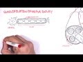
Peripheral nerves have the ability to regenerate. The injured nerve fiber (Axon) is very long and it has to regenerate and reach its target in a reasonable time so that the patient can have a good function. The nerve has protective layers of tissue surrounding it:
•Epineurium: surrounds the nerve itself •Perineurium:L around the fascicle •Fascicle •Nerve fiber •Endoneurium: around the axon •Myelin sheath •Axon
The neurons become stimulated at the dendrites. Neurons possess structures that allow for the transmission of impulses and are composed of two parts:
•Receiving structures: dendrites, cell body •Conducting structures: axon, axon terminal.
Signals from the brain are passed on to the muscles in the limbs by these motor neurons.
When painful stimulus is applied to a sensory receptor in the skin (afferent), the information is transmitted to the central nervous system (CNS). Once the sensory signal has been received by the CNS, another signal is then sent from the brain (efferent) and passed along the motor neurons in order to move the muscles in response to painful stimulus.
When the nerve is affected or cut, there will be no function of that nerve. When the axon is separated from the body, there will be degeneration of the axon and the degeneration will stop at the synapse and will not travel to the next neuron. Regeneration of the peripheral nerves is possible and all events of regeneration occur around the axon when the nerve is cut. The proximal stump will regenerate and the distal stump will have Wallerian degeneration. Macrophagocytes dispose of the degenerated axon and myelin sheath. Schwann cells grew into the cut area and join the two ends. The entire axonal material is phagocytized from the site of the injury to the endplate. Anew generated axon sprouts and grow to reestablish the connection from reaching its target.
•Wallerian degeneration: the neuron is able to survive and regenerate after the axon has been cut with neuronal survival and Wallerian degeneration. The Wallerian degeneration typically arises from severe nerve injuries such as axonotmesis or neurotmesis. The cell body increases in size with migration of the nucleus towards the cell periphery. The cell body enlarges for approximately 20 days and remains enlarged until axon regeneration is complete. In the proximal part of the nerve segment, degeneration can occur and it is proportionated to the severity of the injury. Degeneration extends proximally to the next node of Ranvier. Wallerian degeneration is seen in the distal portion of the nerve fiber.
•Synthesis of growth-promoting molecules: axonal degeneration is followed by degradation of the myelin sheath and infiltration by macrophages.the macrophages are accompanied by Schwann cells which clear the debris from degeneration. Distally you will find Schwann cells proliferate and the axon sprouts (finger like growths) and advance. Using Schwann cells as a guide, these sprouts advance about 1 mm per day. Atrophy of the associated muscle will be seen during the process.
•Regeneration & remyelination: axonal growth is seen and the connection is reestablished with the muscle appearing to be bigger and a bit healthier. When the axon fails to establish continuity, a neuroma formation will be seen with no regeneration of the axon distally. Atrophy of the associated muscle will be seen during the process.
There are three types of nerve injuries:
•Type I: neurapraxia: prognosis is good with neurapraxia and it’s the mildest form of nerve injury and the nerve remains intact.
•Type II: axonotmesis: injury is severe. The axon is damaged and the surrounding connective tissue is intact. There will be partial or complete recovery of the nerve and Wallerian degeneration occurs distal to the nerve injury site. Recovery occurs 1 mm per day or 1 inch per month.
•Type III: neurotmesis: no recovery, fibrillation is present and the injury usually requires surgery. Motor neuron unit potential is usually absent. In neurotmesis there will be degradation and neuroma formation.
EMG and nerve injury
•Degeneration (Wallerian): fibrillation
•Reinnervation(Axon sprouting re-establishment of the connection): polyphasic motor activity is good.
There are several factors that affect the success of recovery. If the gap between the proximal and distal stumps is too wide or scar tissue has formed, surgery can help to guide the sprouts to the tube.
Factors affecting favourable nerve recovery after repair:
•Younger age of the patient •Distal injury •No significant delay in repair •Sharp cuts are better than a crush •Vascularity is preserved •Favorable orientation of the nerve in epineural repair.
Become a friend on facebook:
http://www.facebook.com/drebraheim
Follow me on twitter:
https://twitter.com/#!/DrEbraheim_UTMC
Видео Nerve Regeneration - Everything You Need To Know - Dr. Nabil Ebraheim канала nabil ebraheim
Показать
Комментарии отсутствуют
Информация о видео
Другие видео канала
 Neurology - Nerve Damage and Regeneration
 Can we repair injured nerves? | Massimo Hilliard | TEDxUQ
 Exploring The World of Nerve Repair: Mario Romero at TEDxUTA
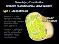 Nerve Injury ,types . Nerve recovery - Everything You Need To Know - Dr. Nabil Ebraheim
 Understanding Nutritional Support for TBI
 Nerve injury , Injuries .Complete - Everything You Need To Know - Dr. Nabil Ebraheim
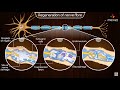 Neuroregeneration in the peripheral nervous system (PNS) - Physiology medical animations
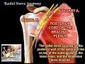 Radial Nerve Anatomy - Everything You Need To Know - Dr. Nabil Ebraheim
 Neurology | Nerve Injury & Repair: Wallerian Degeneration & Regeneration
 Mistakes to avoid if you have nerve damage
 Nerve Regeneration | Spinal Cord vs Peripheral Nerves
 Nerve Regeneration | Wallerian Degeneration | Nerve Damage | ANIMATION | Neuron | The Young Orthopod
 Spinal Cord Injury, Motor Neurons, and Reflexes
 Nerve Regeneration - Restore Nerve Connections / Repair Nerve Growth - Binaural Beats Meditation
 Compression of the Spinal Cord & Hand Function - Everything You Need To Know - Dr. Nabil Ebraheim
 Nerve repair: Regeneration in spinal-cord injury
 Why Can't We Reverse Nerve Damage?
 Is My Nerve Pain Getting Worse... or Better? | The Nerve Behavior Scale
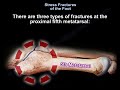 Stress Fractures of the Foot - Everything You Need To Know - Dr. Nabil Ebraheim
 Binaural Beats - Nerve and Cell Regeneration Meditation Tone