Neuroregeneration in the peripheral nervous system (PNS) - Physiology medical animations
►𝐉𝐨𝐢𝐧 𝐓𝐡𝐢𝐬 𝐂𝐡𝐚𝐧𝐧𝐞𝐥 𝐓𝐨 𝐆𝐞𝐭 𝐀𝐜𝐜𝐞𝐬𝐬 𝐓𝐨 𝐏𝐞𝐫𝐤𝐬 :- https://bit.ly/2RQHvTN
►𝐃𝐨𝐰𝐧𝐥𝐨𝐚𝐝 𝐭𝐡𝐞 𝐌𝐞𝐝𝐯𝐢𝐳𝐳 𝐚𝐩𝐩 𝐮𝐬𝐢𝐧𝐠 𝐭𝐡𝐞 𝐛𝐞𝐥𝐨𝐰 𝐥𝐢𝐧𝐤 👇👇👇👇 𝐃𝐨𝐰𝐧𝐥𝐨𝐚𝐝 👇👇👇👇
►𝐀𝐧𝐝𝐫𝐨𝐢𝐝 :- https://bit.ly/3ansFKq
📌𝐅𝐨𝐥𝐥𝐨𝐰 𝐨𝐧 𝐈𝐧𝐬𝐭𝐚𝐠𝐫𝐚𝐦 :-
https://www.instagram.com/drgbhanuprakash
Peripheral nervous system regeneration
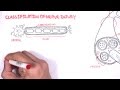
Neuroregeneration in the peripheral nervous system (PNS) occurs to a significant degree.Axonal sprouts form at the proximal stump and grow until they enter the distal stump. The growth of the sprouts is governed by chemotactic factors secreted from Schwann cells (neurolemmocytes). Injury to the peripheral nervous system immediately elicits the migration of phagocytes, Schwann cells, and macrophages to the lesion site in order to clear away debris such as damaged tissue. When a nerve axon is severed, the end still attached to the cell body is labeled the proximal segment, while the other end is called the distal segment. After injury, the proximal end swells and experiences some retrograde degeneration, but once the debris is cleared, it begins to sprout axons and the presence of growth cones can be detected. The proximal axons are able to regrow as long as the cell body is intact, and they have made contact with the Schwann cells in the endoneurial channel or tube. Human axon growth rates can reach 1 mm/day in small nerves and 5 mm/day in large nerves.The distal segment, however, experiences Wallerian degeneration within hours of the injury; the axons and myelin degenerate, but the endoneurium remains. In the later stages of regeneration the remaining endoneurial tube directs axon growth back to the correct targets. During Wallerian degeneration, Schwann cells grow in ordered columns along the endoneurial tube, creating a band of Büngner (boB) that protects and preserves the endoneurial channel. Also, macrophages and Schwann cells release neurotrophic factors that enhance re-growth.
#physiologyvideos #usmlevideos #physiologyofpns #usmlestep1videos #animatedmedicalvideos #physologyanimatedlectures #drgbhanuprakash #mbbs #neetpg #fmge #pnsphysiology #walleriandegeneration #regenerationofPNS #regenerationofnervoussystem #regenerationofneuron #regenerationofperipheralnervoussystem #nervoussystemregeneration #physiology #medicalanimations
Видео Neuroregeneration in the peripheral nervous system (PNS) - Physiology medical animations канала Dr.G Bhanu Prakash Animated Medical Videos
►𝐃𝐨𝐰𝐧𝐥𝐨𝐚𝐝 𝐭𝐡𝐞 𝐌𝐞𝐝𝐯𝐢𝐳𝐳 𝐚𝐩𝐩 𝐮𝐬𝐢𝐧𝐠 𝐭𝐡𝐞 𝐛𝐞𝐥𝐨𝐰 𝐥𝐢𝐧𝐤 👇👇👇👇 𝐃𝐨𝐰𝐧𝐥𝐨𝐚𝐝 👇👇👇👇
►𝐀𝐧𝐝𝐫𝐨𝐢𝐝 :- https://bit.ly/3ansFKq
📌𝐅𝐨𝐥𝐥𝐨𝐰 𝐨𝐧 𝐈𝐧𝐬𝐭𝐚𝐠𝐫𝐚𝐦 :-
https://www.instagram.com/drgbhanuprakash
Peripheral nervous system regeneration
Neuroregeneration in the peripheral nervous system (PNS) occurs to a significant degree.Axonal sprouts form at the proximal stump and grow until they enter the distal stump. The growth of the sprouts is governed by chemotactic factors secreted from Schwann cells (neurolemmocytes). Injury to the peripheral nervous system immediately elicits the migration of phagocytes, Schwann cells, and macrophages to the lesion site in order to clear away debris such as damaged tissue. When a nerve axon is severed, the end still attached to the cell body is labeled the proximal segment, while the other end is called the distal segment. After injury, the proximal end swells and experiences some retrograde degeneration, but once the debris is cleared, it begins to sprout axons and the presence of growth cones can be detected. The proximal axons are able to regrow as long as the cell body is intact, and they have made contact with the Schwann cells in the endoneurial channel or tube. Human axon growth rates can reach 1 mm/day in small nerves and 5 mm/day in large nerves.The distal segment, however, experiences Wallerian degeneration within hours of the injury; the axons and myelin degenerate, but the endoneurium remains. In the later stages of regeneration the remaining endoneurial tube directs axon growth back to the correct targets. During Wallerian degeneration, Schwann cells grow in ordered columns along the endoneurial tube, creating a band of Büngner (boB) that protects and preserves the endoneurial channel. Also, macrophages and Schwann cells release neurotrophic factors that enhance re-growth.
#physiologyvideos #usmlevideos #physiologyofpns #usmlestep1videos #animatedmedicalvideos #physologyanimatedlectures #drgbhanuprakash #mbbs #neetpg #fmge #pnsphysiology #walleriandegeneration #regenerationofPNS #regenerationofnervoussystem #regenerationofneuron #regenerationofperipheralnervoussystem #nervoussystemregeneration #physiology #medicalanimations
Видео Neuroregeneration in the peripheral nervous system (PNS) - Physiology medical animations канала Dr.G Bhanu Prakash Animated Medical Videos
Показать
Комментарии отсутствуют
Информация о видео
7 марта 2018 г. 16:44:11
00:02:53
Другие видео канала
 Neurology - Nerve Damage and Regeneration
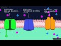 Action Potential in the Neuron
 You can grow new brain cells. Here's how | Sandrine Thuret
 Saltatory conduction - Conduction through Myelinated nerve fiber : Physiology medical animations
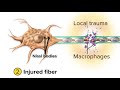 Damage and Repair in the Peripheral Nervous System
 Nerve Injury ,types . Nerve recovery - Everything You Need To Know - Dr. Nabil Ebraheim
 Neurology | Gross Anatomy of the Spinal Cord and Spinal Nerves
 Regenerative Medicine as Neuropathy Treatment with Dr. Sebastian in Estero FL
 Peripheral Nervous System: Crash Course A&P #12
 Neurology | Olfactory Nerve: Cranial Nerve I
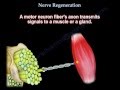 Nerve Regeneration - Everything You Need To Know - Dr. Nabil Ebraheim
 Neurology | Nerve Injury & Repair: Wallerian Degeneration & Regeneration
 Nerve repair: Regeneration in spinal-cord injury
 Why Can't We Reverse Nerve Damage?
 Wallerian degenration and regeneration | Nerve injury classification | Physiology mbbs 1st year
 Overview of the Peripheral Nervous System
 Nerve Regeneration | Wallerian Degeneration | Nerve Damage | ANIMATION | Neuron | The Young Orthopod
 NEUROANATOMY-DEVELOPMENT OF THE NERVOUS SYSTEM-PART-1-NEURULATION-DR ROSE JOSE MD
 Nerve Injury | Nerve Damage | ANIMATION | Neuron | The Young Orthopod
 Parkinson's Disease (Shaking Palsy) - Clinical Presentation and Pathophysiology