Fundamentals of Slit Lamp Biomicroscopy
[MUSIC PLAYING] SPEAKER: Slit lamp biomicroscopy is an acquired skill. This introduction will help you over the initial hurdles that all face when confronted with their first patient at the slit lamp. The purpose of this video is to acquaint you with all the features and uses of slit lamp biomicroscopy.
With this information, you can begin concentrating on what you're seeing rather than thinking about the mechanics of the examination. As Yogi Berra says, you can see a lot just by observing. Slit lamp biomicroscopy has four advantages over loop observations, better magnification, improved stereopsis, superior illumination, and excellent depth localization.
The slit lamp produces a very bright and focused beam of light that can be varied from a hairline slit to an 8-millimeter wide beam or to a small spot beam of light. The light source is coupled by a common carriage to a binocular microscope observation system. Both light and magnifier are parfocal, that is, focused at the exact same place.
Therefore, when the slit beam is seen in sharp focus, the object on which it is focused will be clearly seen through the oculars. Fine manipulations of the slit beam can then aid in evaluating an area of interest or abnormality, such as this corneal scar. The slit beam can pivot independently, side to side, through 180 degrees to permit viewing of any ocular structure from different angles, particularly helpful in depth localization.
The basic slip lamp biomicroscopy exam procedure is as follows. 1, adjust the headrest and height of the chin rest for the patient's maximum comfort, and proper viewing level. The patient's eye should be level with the black line on the headrest column. This indicates there will be equal vertical maneuvering room above and below this line. 2, set the eyepieces at 0, or for your refractive error, and adjust the inner pupillary distance of the oculars.
3, turn on the power to the slit lamp. 4, release the fixing screw that locks the base or cross slide. 5, grasp the base carriage or cross slide with both hands and move it so the slit beam is focused on the patient's temporal or lateral conjunctiva. This is done with the naked eye and not by looking through the oculars.
Now, look through the microscope and fine focus the slit beam by maneuvering the joystick. Note that the light beam can be elevated or lowered by twisting or rotating the joystick. To vary the dimensions of the light, the following controls must be adjusted. Slit beam length is manipulated by turning the upper control lever knob. The lever also controls slit beam orientation or rotation.
Slit being width is controlled by turning the lower control knobs. Beam intensity and color are controlled by the transformer output and upper filter lever. Increasing the voltage from the standard 5 to 7 and 1/2 volts will double the brightness, but it'll also significantly shorten the life of the lamp...
...The Haag-Streit slit beam can be inclined or tilted by depressing the latch at the base of the lamp arm. By tilting, rotating, and coaxially setting the slit beam to horizontal and using the Goldmann 3 mirror fundis lens, the peripheral vitreous and retina exam can be optimized. Utilizing the tilt feature with a horizontal slit beam can eliminate unwanted and interfering light reflections in all methods of fundis biomicroscopy.
In conclusion, the dynamic use of the slit beam size, orientation, and its relative position with a microscope will produce the most information in your examinations. Careful slit lamp biomicroscopy will reward you with a great deal of valuable information, information that can be essential to the proper diagnosis and management of many ocular diseases. This introduction to slit lamp biomicroscopy should make your early efforts easier and more successful so that you may become more accomplished and provide better patient care.
[MUSIC PLAYING]
Видео Fundamentals of Slit Lamp Biomicroscopy канала UVA Health - School of Medicine
Показать
Комментарии отсутствуют
Информация о видео
13 января 2021 г. 0:04:02
00:23:32
Другие видео канала
 Retina exam on slit lamp - COMPLETE GUIDE
 Ophthalmic Skills Series Part 1/5
 How to Use a Slit Lamp | Explained by an Ophthalmologist
 Slit Lamp Techniques Optical Section of Crystalline Lens
 Slit Lamp Techniques - OPHTHALMOLOGY - Ep 2
 Part 3: Using a Slit Lamp
 Direct Ophthalmoscope - OPHTHALMOLOGY - Ep 3
 Slit Lamp Examination (Biomicroscopy) in English
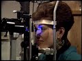 Goldmann Applanation Tonometry
 Slit Lamp Overview - OPHTHALMOLOGY - Ep 1
 Slit Lamp Techniques Van Herick's
 Slit Lamp
 Slit Lamp Exam
 Approach to Fundoscopy / Ophthalmoscopy
 The Slit Lamp Exam – Episode 1, Components of the Slit Lamp
 Slit Lamp Techniques Retro Illumination
 Slit lamp Examination Techniques
 Slit Lamp Exam Tutorial - Part 1
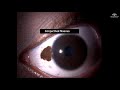 Slit Lamp examination techniques for beginners
 Learning the Ophthalmoscope