Goldmann Applanation Tonometry
A fundamental component of every eye examination is a determination of intraocular pressure. This assessment provides information on the ordinary function of the eye and assists in the detection of glaucoma. Once you understand the principles, the mechanics of checking intraocular pressure are straightforward.
In this videotape, we will review how to measure intraocular pressure by Goldmann Applanation Tonometry. An individual's intraocular pressure may vary with time of day, posture, exertion, lid movement, medications, and diet. A single reading is only an estimate of the fluctuations that occur.
From general population studies, it has been determined that approximately 95% of the population has an intraocular pressure of between 6 and 21 millimeters of mercury. There are a number of factors which have an effect on average intraocular pressure. These include race, refractive error, and age.
Elevated intraocular pressure is not limited to the elderly. Glaucoma can occur at any age. Therefore, every patient who is able or willing to cooperate should have intraocular pressure checked regularly. The average intraocular pressure is about 15 millimeters of mercury.
Hypotony refers to an intraocular pressure below 6 millimeter of mercury. Hypotony for prolonged periods of time is associated with deleterious effects on the eye. Intraocular pressures above 21 millimeter of mercury increase the suspicion for glaucoma. The higher the intraocular pressure, the greater the likelihood of developing damage to the eye.
The optic nerve is most susceptible to this elevation in pressure. A patient with elevated intraocular pressure but no damage to the visual field or optic nerve is considered a glaucoma suspect or ocular hypertensive. If damage in the nerve or visual field is detected, a diagnosis of glaucoma is made.
There are a variety of familiar ways to measure the pressure of an object, such as by palpation or by inserting a manometer or gauge. Since this cannot be done in a human eye, indirect methods of tonometry are used. Applanation tonometers measure the force necessary to flatten a corneal surface of constant size.
We will concentrate on applanation tonometry with a Goldmann tonometer since it is generally considered to be the standard to which others are compared. It is attached to the slit lamp biomicroscope and consists of an applanating replaceable tip containing a plastic biprism attached by a rod to the housing piece inside, which contains a coiled spring and a series of levers that are used to adjust the force of the biprism against the cornea. Two beams splitting prisms within the operating unit optically convert the circular area of corneal contact into two semicircles.
An important component of applanation tonometry is the tear film meniscus. The tear film meniscus is stained with fluorescein to allow visualization at the margin of contact between the cornea and biprism. As already noted, the circular area of contact is converted by the prisms into two semicircles called mires, seen here through the slit lamp.
The force of touch or applanation is adjusted until the inner edges overlap. Intraocular pressure is then read directly from a scale on the tonometer housing. The marks on the scale are in 2 millimeters of mercury increments, although intraocular pressure can be estimated to the nearest 1 millimeter. The scale number is multiplied by 10 and this corresponds directly to the intraocular pressure in millimeters of mercury.
In performing applanation tonometry, the patient should be seated comfortably at the slit lamp. The chin and forehead should be in their proper rests, a tight fitting collar should be loosened. There may be difficulty positioning some patients. It helps in these settings to have the patient lean forward in the chair.
The patient's eyes should be aligned with this marker on the slit lamp coil. After the slit lamp is turned on to its highest level of illumination, the cobalt blue filter is put into place and the slit open to its widest position to achieve the brightest light. The angle between the illumination device and the microscope should be about 60 degrees to best illuminate the tip of the prism....
...The necessary equipment and instructions for this are included with the applanation tonometer. If proper calibration cannot be achieved, the transmitter should be sent to a service center or the manufacturer. In summary, Goldmann applanation tonometry is an important part of a complete eye examination. Developing a routine of testing is essential. Proper positioning of the patient at the slit lamp, maximum brightness of the spot, and good technique will ensure accuracy and reliability for all patients.
Видео Goldmann Applanation Tonometry канала UVA Health - School of Medicine
Показать
Комментарии отсутствуют
Информация о видео
13 января 2021 г. 0:01:40
00:12:51
Другие видео канала
 The Ophthalmic Exam: Retina and Posterior Segment
 OT skills guide: Applanation tonometry
 Goldmann applanation Tonometry (GAT) - basic STEP BY STEP GUIDE
 Ophthalmology Made Ridiculously Easy | 1st Edition | Digital Book
 A brief guide to gonioscopy
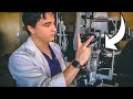 How to Use a Slit Lamp | Explained by an Ophthalmologist
 User Guide for the Icare Tonometer
 Macular OCT Interpretation: A Practical Discussion with Dr. David E. Lederer
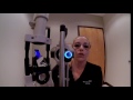 First Person Routine Eye Exam
 Using Subjective Refraction to Calculate Glasses Prescription and Fit a Contact Lens
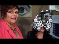 Subjective Refractometry Technique for Ophthalmic Technicians (Eye Techs)
 Ophthalmic Skills Series Part 1/5
 Natural Glaucoma Treatment for High Eye Pressure - How to Lower Eye Pressure Naturally
 ASMR: Relaxing Glaucoma Tests ft. The Perkins Tonometer! (roleplay)
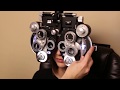 How to Perform a Manifest Refraction
 An introduction to Goldmann Applanation Tonometry
 Eye tonometry
 The Slit Lamp Exam – Episode 5, Retro Illumination
 Goldmann Applanation Tonometry- Theory and Practice Edition II | Dr. M. D. Singh
 Dr. Swathi Reddy "What Eye Pressure is Right for Me?" GSF CARES Conference