Fracture Healing - Everything You Need To Know - Dr. Nabil Ebraheim
Dr. Ebraheim's animated educational video describing fracture healing.
The stability of the fracture decides what type of healing will occur. If there is a small amount of strain (below 2%), the primary bone healing will occur. The primary bone healing will occur like when you are using a compression plate. If the strain is between 2-10%, then secondary bone healing will occur such as when using a cast, rod or external fixator. In primary bone healing, you will need absolute stability, called Haversian remodeling cutting cone remodeling, or sometimes called intramembranous healing.
Secondary bone healing will occur when the fixation is not rigid, such as with a cast, and there will be endochondral ossification. With an IM rod, there will be secondary bone healing, early on, there will be periosteal callus. Later on, there will be a medullary callus.
The external fixator is predominantly periosteal callus with endochondral ossification, because most of the time the external fixator is not very rigid. When endochondral ossification fails, because the fixation is inadequate, you get hypertrophic nonunion and you will have predominantly type II collagen. The endochondral ossification at this point has failed and stability is needed in order to change the cartilage to bone.
What are the stages of fracture healing?
1-Hematoma
2-Inflammation
3-Soft callus
4-Hard callus
5-Remodeling
When the fracture occurs, there will be bleeding at the fracture site. It will make a fibrin blood clot.
1-Stage of inflammation
Cells
•Macrophages
•Mesenchymal cells
•Stem cells migrate to the fracture and form the granulation tissue and will release the growth factors. Granulation tissue tolerates the greatest strain before failure.
COX-2 inhibitor and nonsteroidal depresses Runx2. Important for the differentiation of osteoblasts.
2-Stage of soft callus
•Will occur within two weeks.
•The amount of callus correlates with the immobilization.
•The stiffer the immobilization, the less amount of callus.
•Flexible fixation will result in endochondral ossification (abundant callus).
•Secondary bone healing: healing through cartilage formation. The stability helps direct bone formation. Lack of stability helps the formation of cartilage, which later on can change to endochondral ossification.
3-Stage of hard callus
•The collagen changes from predominantly type II, to be followed by type I.
•Type I= bone
•Type II =cartilage
4-Stage of remodeling
•The stage of remodeling will begin at 2 weeks and continue many years after the fracture has healed.
•The woven bone will be replaced by stronger, laminar bone and the fracture healing will be complete with the continuation of the medullary cavity.
•The remodeling of the bone is influenced by the Wolff’s law (means that the bone is affected by stress).
You need to be aware of this order of bone healing because sometimes it comes on the exam.
Blood flow at the fracture site is very important for fracture healing. The blood will supply the fracture with nutrients and cells. Initially there Is decreased blood flow at the fracture site which will increase later on and the blood flow will peak at two weeks and return to normal after about three months.
Distraction osteogenesis
Can get type I and type II cartilage (predominantly type I) because there is more intramembranous ossification.
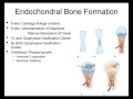
How does the endochondral bone formation occur?
•Chondrocyte proliferation then hypertrophy
•Matrix mineralization
•The chondrocytes die
•Vascular invasion, ossification, and remodeling to lamellar bone.
What are the growth factors involved in fracture healing?
Bone morphogenic protein: osteoinductive causing the undifferentiated mesenchymal cells to differentiate into osteoblasts.
Transforming growth factor beta (TGF-B1): will make the mesenchymal cells produce type II collagen and proteoglycans (trying to produce endochondral ossification)
Insulin-like growth factor 2 (IGF-2): will stimulate type I collagen
Platelet derived growth factor (PDGF): will be released from the platelets. Attracts inflammatory cells to the fracture site (chemotactic).
Follow me on twitter:
https://twitter.com/#!/DrEbraheim_UTMC
Donate to the University of Toledo Foundation Department of Orthopaedic Surgery Endowed Chair Fund:
https://www.utfoundation.org/foundation/home/Give_Online.aspx?sig=29
Видео Fracture Healing - Everything You Need To Know - Dr. Nabil Ebraheim канала nabil ebraheim
The stability of the fracture decides what type of healing will occur. If there is a small amount of strain (below 2%), the primary bone healing will occur. The primary bone healing will occur like when you are using a compression plate. If the strain is between 2-10%, then secondary bone healing will occur such as when using a cast, rod or external fixator. In primary bone healing, you will need absolute stability, called Haversian remodeling cutting cone remodeling, or sometimes called intramembranous healing.
Secondary bone healing will occur when the fixation is not rigid, such as with a cast, and there will be endochondral ossification. With an IM rod, there will be secondary bone healing, early on, there will be periosteal callus. Later on, there will be a medullary callus.
The external fixator is predominantly periosteal callus with endochondral ossification, because most of the time the external fixator is not very rigid. When endochondral ossification fails, because the fixation is inadequate, you get hypertrophic nonunion and you will have predominantly type II collagen. The endochondral ossification at this point has failed and stability is needed in order to change the cartilage to bone.
What are the stages of fracture healing?
1-Hematoma
2-Inflammation
3-Soft callus
4-Hard callus
5-Remodeling
When the fracture occurs, there will be bleeding at the fracture site. It will make a fibrin blood clot.
1-Stage of inflammation
Cells
•Macrophages
•Mesenchymal cells
•Stem cells migrate to the fracture and form the granulation tissue and will release the growth factors. Granulation tissue tolerates the greatest strain before failure.
COX-2 inhibitor and nonsteroidal depresses Runx2. Important for the differentiation of osteoblasts.
2-Stage of soft callus
•Will occur within two weeks.
•The amount of callus correlates with the immobilization.
•The stiffer the immobilization, the less amount of callus.
•Flexible fixation will result in endochondral ossification (abundant callus).
•Secondary bone healing: healing through cartilage formation. The stability helps direct bone formation. Lack of stability helps the formation of cartilage, which later on can change to endochondral ossification.
3-Stage of hard callus
•The collagen changes from predominantly type II, to be followed by type I.
•Type I= bone
•Type II =cartilage
4-Stage of remodeling
•The stage of remodeling will begin at 2 weeks and continue many years after the fracture has healed.
•The woven bone will be replaced by stronger, laminar bone and the fracture healing will be complete with the continuation of the medullary cavity.
•The remodeling of the bone is influenced by the Wolff’s law (means that the bone is affected by stress).
You need to be aware of this order of bone healing because sometimes it comes on the exam.
Blood flow at the fracture site is very important for fracture healing. The blood will supply the fracture with nutrients and cells. Initially there Is decreased blood flow at the fracture site which will increase later on and the blood flow will peak at two weeks and return to normal after about three months.
Distraction osteogenesis
Can get type I and type II cartilage (predominantly type I) because there is more intramembranous ossification.
How does the endochondral bone formation occur?
•Chondrocyte proliferation then hypertrophy
•Matrix mineralization
•The chondrocytes die
•Vascular invasion, ossification, and remodeling to lamellar bone.
What are the growth factors involved in fracture healing?
Bone morphogenic protein: osteoinductive causing the undifferentiated mesenchymal cells to differentiate into osteoblasts.
Transforming growth factor beta (TGF-B1): will make the mesenchymal cells produce type II collagen and proteoglycans (trying to produce endochondral ossification)
Insulin-like growth factor 2 (IGF-2): will stimulate type I collagen
Platelet derived growth factor (PDGF): will be released from the platelets. Attracts inflammatory cells to the fracture site (chemotactic).
Follow me on twitter:
https://twitter.com/#!/DrEbraheim_UTMC
Donate to the University of Toledo Foundation Department of Orthopaedic Surgery Endowed Chair Fund:
https://www.utfoundation.org/foundation/home/Give_Online.aspx?sig=29
Видео Fracture Healing - Everything You Need To Know - Dr. Nabil Ebraheim канала nabil ebraheim
Показать
Комментарии отсутствуют
Информация о видео
Другие видео канала
 Fracture healing and repair pt 1 - basic science
 Fracture Healing | ANIMATION | BASICS | The Young Orthopod
 Hip Fractures - Everything You Need To Know - Dr. Nabil Ebraheim
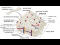 Bones: Structure and Types
 Humerus Fractures - Everything You Need To Know - Dr. Nabil Ebraheim
 Bone Fracture - Types, Fracture Repair and Osteomyelitis
 Bone Fracture: Types & Mechanisms | ANIMATION | Fracture classification | The Young Orthopod NEET PG
 Humeral Shaft Fractures Treatment Alternative - Everything You Need To Know - Dr. Nabil Ebraheim
 Common Types Of Distal Radius Fractures - Everything You Need To Know - Dr. Nabil Ebraheim
 Fracture Healing Part 2 - Everything You Need to Know - Dr. Nabil Ebraheim
 The Wonders of Bone Healing
 Bone Fractures Types Nursing Interventions, Treatment, Signs and Symptoms NCLEX
 How Does a Bone Heal?
 MUST Do Exercises with Injured Foot or Ankle- Faster Recovery
 Metacarpal Fractures - Everything You Need To Know - Dr. Nabil Ebraheim
 Fractures Of The Femur Shaft Winquist & Hansen - Everything You Need To Know - Dr. Nabil Ebraheim
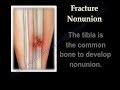 NONUNION OF FRACTURES CAUSES AND TREATMENT - Everything You Need To Know - Dr. Nabil Ebraheim
 Fracture healing
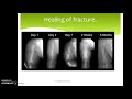 Wound healing and repair (part 2)- Fracture healing
 Low Back Pain - Everything You Need To Know - Dr. Nabil Ebraheim