60 Seconds of Echo Teaching Answer: A left atrial appendage thrombus?
This is an example of a thrombus in formation in the left atrial appendage. Not yet a thrombus though. Take a look and share your thoughts in the comment section below.
Learn more with 123sonography, the global #1 online platform for learning medical ultrasound & echocardiography. 123sonography offers the right product - all 100% online. Register for free chapters on our website today! https://www.123sonography.com/ultrasound-and-echocardiography
While developing the courses and defining the learning objectives at 123sonography, we strictly follow the scientific guidelines on how to assess and manage your patients with the help of ultrasound. Choose between Diploma Courses, micro-learning apps, webinars, and subscription memberships – we have a matching learning path ready for you!
#Ultrasound #Echocardiography #eLearning
Видео 60 Seconds of Echo Teaching Answer: A left atrial appendage thrombus? канала 123sonography
Показать
Комментарии отсутствуют
Информация о видео
Другие видео канала
 Detection of Left Atrial Thrombus - A/Prof Phillip Mottram
 60 Seconds of Echo Teaching Answer: Does this patient have HCMP?
 60 Seconds of Echo Teaching Answer: Is this a myxoma?
 TEE Essentials: Mastering the mid-esophageal left atrial appendage view
 60 Seconds of Echo Teaching Answer: STEMI - RCA or CX?
 60 Seconds of Echo Teaching Answer: A congenital defect?
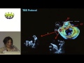 Bushra Rana - The gold standard; TEE assessment before, during, and after LAA occlusion
 Diastolic Function — A Simple Approach
 Transesophageal Echocardiography : Left Atrial Appendage Thrombus
 60 Seconds of Echo Teaching Answer: Which procedure was performed on this patient?
 EACVI free webinar: How to use contrast to rule out left atrial appendage thrombus
 Left atrial appendage closure - a case based discussion
 Point of Care Echo: Stroke Volume Determination
 Ischemic Cardiomyopathy: Entire Echo Exam
 What can I find when I use color Doppler in a PLAX?
 60 Seconds of Echo Teaching Answer: A strange structure.
 Assessment of Masses in Echocardiography
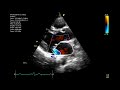 Echocardiographic assessment of the mitral valve
 An overview of Atrial Fibrillation
 Echo Teaching Challenge Answer: What problem is present in this patient?