60 Seconds of Echo Teaching Answer: A congenital defect?
Here is the answer to our latest question. The patient not only has a persistent left superior vena cava but an upper sinus venous defect as well. The dilated right ventricle and the high flow (VTI) across the pulmonary artery indicated that there is a shunt.
LET US BE YOUR MENTOR. EASY SELF-PACED LEARNING METHOD:
No matter what your level of expertise is, if you work in an emergency setting, in general medicine, primary care or if you are training to become a doctor or sonographer, we have a matching learning path ready for you. 123sonography offers the right product - all 100% online. Register for free chapters here: https://www.123sonography.com/
We help you to get started in ultrasound and echocardiography with our Diploma BachelorClasses. With our Diploma MasterClasses you will be able to deepen your existing knowledge and become a master in the field. For those of you already skilled in echo, and wish to stay up to date, we offer continuous micro-learning knowledge training with our Premium Memberships, Apps, Quizzes and Webinars. While developing the courses and defining the learning objectives at 123sonography, we strictly follow the scientific guidelines on how to assess and manage your patients with the help of ultrasound.
Best regards,
123sonography
Видео 60 Seconds of Echo Teaching Answer: A congenital defect? канала 123sonography
Показать
Комментарии отсутствуют
Информация о видео
Другие видео канала
 What problems are there with ejection fraction?
 Dilated coronary sinus due to significant pulmonary hypertension
 Ultrasound Case 140 - Eagle's Syndrome
 Is Marrying Your Cousin Actually Dangerous?
 Persistent Left Superior Vena Cava (PLSVC)
 Embryology for the Cardiologist (Regina Lantin-Hermoso, MD) May 3, 2016
 Adult Congenital Heart Disease: Anomalous Coronary Arteries (Risk Stratification)
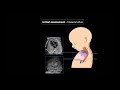 Fetal Echocardiography: Protocol and Technique
 Dilated Cardiomyopathy - causes, symptoms, pathophysiology and treatment
 What can I find when I use color Doppler in a PLAX?
 Clinical Anatomy - Cardiac Coronary Vessels (left and right coronary artery, venous sinus)
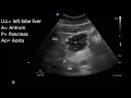 Gastric ultrasound for preoperative assessment
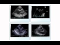 Native Valve Endocarditis
 How to obtain: CRAB VIEW (Paediatric Echocardiography)!
 Persistent Left Superior Vena Cava (PLSVC)
 Superior vena cava and the azygos system clinical anatomy - SVC obstruction (oncology emergency)
 Point-of-Care Echo: Regional Wall Motion Abnormalities
 Hot Tips - IVC Volume Assessment with Ultrasound
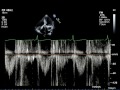 Echo Challenge: Mitral Regurgitation
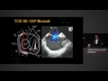 Atrial Septum - Anatomy, Echocardiography & Interventions - A/Prof Gregory Scalia