Histology of Parathyroid Gland
Link of My Instagram : https://instagram.com/knowing_anatomy?igshid=12mmn11n2pxe6
The parathyroid glands are four nodular structures, typically located on the dorsum of the thyroid at each of its four poles. These glands monitor the serum calcium level and secrete parathyroid hormone (PTH) when it is low. PTH is essential for maintaining calcium homeostasis. Thus, dysregulation of this hormone can lead to various pathologies. Histological examination is an important technique used to evaluate and diagnose these pathologies, such as primary, secondary, and tertiary hyperparathyroidism and parathyroid carcinoma.
The parathyroid glands are nodular configurations derived from endodermal tissue on the dorsum of the thyroid gland. Typically, four of these structures are present with the superior parathyroids located at the upper poles and inferior parathyroids at the lower poles of the thyroid; however, the number and location of these glands are variable. Parathyroid glands may occasionally be found ectopically within the mediastinum and may be present in numbers greater or fewer than four.The superior parathyroids are frequently located close to the cricothyroid junction, above the intersection between the inferior thyroid artery and recurrent laryngeal nerve.The location of the inferior parathyroid glands is much less consistent. The inferior thyroid artery most commonly supplies the parathyroid glands; however, their blood supply may also come from the superior thyroid artery. The glandular tissue of the parathyroids is separated from that of the thyroid by a fibrous capsule. The parenchyma is primarily composed of two cell types, known as chief and oxyphil cells.
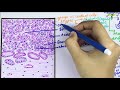
Composed primarily of chief cells and fat with thin fibrous capsule dividing gland into lobules
May have a pseudofollicle pattern resembling thyroid follicles (pink material is PAS positive)
Chief cells:
6 - 8 microns, polygonal, central round nuclei, contain granules of parathyroid hormone (PTH)
Basic cell type, other cell types are due to differences in physiologic activity
80% of chief cells have intracellular fat
Chief cell is most sensitive to changes in ionized calcium
Oxyphil cells:
Slightly larger than chief cell (12 microns), acidophilic cytoplasm due to mitochondria
No secretory granules
First appear at puberty as single cells, then pairs, then nodules at age 40
Water clear cell:
Abundant optically clear cytoplasm and sharply defined cell membranes
Chief cells with excessive cytoplasmic glycogen
Видео Histology of Parathyroid Gland канала Knowing Anatomy
The parathyroid glands are four nodular structures, typically located on the dorsum of the thyroid at each of its four poles. These glands monitor the serum calcium level and secrete parathyroid hormone (PTH) when it is low. PTH is essential for maintaining calcium homeostasis. Thus, dysregulation of this hormone can lead to various pathologies. Histological examination is an important technique used to evaluate and diagnose these pathologies, such as primary, secondary, and tertiary hyperparathyroidism and parathyroid carcinoma.
The parathyroid glands are nodular configurations derived from endodermal tissue on the dorsum of the thyroid gland. Typically, four of these structures are present with the superior parathyroids located at the upper poles and inferior parathyroids at the lower poles of the thyroid; however, the number and location of these glands are variable. Parathyroid glands may occasionally be found ectopically within the mediastinum and may be present in numbers greater or fewer than four.The superior parathyroids are frequently located close to the cricothyroid junction, above the intersection between the inferior thyroid artery and recurrent laryngeal nerve.The location of the inferior parathyroid glands is much less consistent. The inferior thyroid artery most commonly supplies the parathyroid glands; however, their blood supply may also come from the superior thyroid artery. The glandular tissue of the parathyroids is separated from that of the thyroid by a fibrous capsule. The parenchyma is primarily composed of two cell types, known as chief and oxyphil cells.
Composed primarily of chief cells and fat with thin fibrous capsule dividing gland into lobules
May have a pseudofollicle pattern resembling thyroid follicles (pink material is PAS positive)
Chief cells:
6 - 8 microns, polygonal, central round nuclei, contain granules of parathyroid hormone (PTH)
Basic cell type, other cell types are due to differences in physiologic activity
80% of chief cells have intracellular fat
Chief cell is most sensitive to changes in ionized calcium
Oxyphil cells:
Slightly larger than chief cell (12 microns), acidophilic cytoplasm due to mitochondria
No secretory granules
First appear at puberty as single cells, then pairs, then nodules at age 40
Water clear cell:
Abundant optically clear cytoplasm and sharply defined cell membranes
Chief cells with excessive cytoplasmic glycogen
Видео Histology of Parathyroid Gland канала Knowing Anatomy
Показать
Комментарии отсутствуют
Информация о видео
Другие видео канала
 Histology of Pituitary gland \ Hypophysis Cerebri
 Endocrinology | Parathyroid Gland | Calcitonin
 HISTOLOGY OF THYROID GLAND || Epithelium || Follicular cells || Parafollicular cells || Structure |
 Anatomy of Parathyroid Glands - Location , Blood supply , Nerve supply , Histology , Development
 Thyroid Gland Anatomy - (embryology, blood supply, venous drainage, innervation, histology)
 Histology of Hyaline Cartilage
 Shotgun Histology Parathyroid
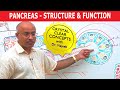 Pancreas - Structure & Function
 Histology of Cardiovascular System
 Histology of Adrenal gland - Shotgun Histology
 Thyroid and Parathyroid Glands
 Liver Histology - Hepatocytes & Portal Vein
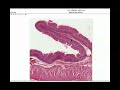 Anatomy | Histology of the Stomach & Small Intestine
 Neurology | Nerve Injury: Wallerian Degeneration & Regeneration
 Parathyroid Hormone Made Easy
 Thyroid Hormones Synthesis - Thyroid Gland
 Histology Helper - Male Reproductive Histology
 Endocrine Histology: Pituitary Gland – Histology | Lecturio
 Histology of thyroid gland - Shotgun Histology
 Parotid Gland - 1 | External Features & Capsule of Parotid