Histology of Hyaline Cartilage
Hyaline cartilage is the most common of the three types of cartilage. In its fresh state, it is homogeneous and semi-transparent. In adults, hyaline cartilage is located in the articular surfaces of movable joints, in the walls of the respiratory tracts (nose, larynx, trachea, and bronchi), in the costal cartilages, and in the epiphyseal plates of long bones.
These locations are easy to remember if you use a mnemonic! In this case 'BLANCET' (read blanket), will help you remember the following structures:
Bronchial cartilage
Laryngeal cartilage
Articular cartilage
Nasal cartilage
Costal cartilage
Epiphyseal growth plates
Tracheal
During embryonic development, hyaline cartilage serves as temporary cartilage models that are essential precursors to the formation of most of the axial and appendicular skeleton. This article will focus on important features of hyaline cartilage, namely its matrix, chondrocytes, and perichondrium.
Chondrocytes
Chondrocytes occupy relatively little of the hyaline cartilage mass. They are embedded in an extensive matrix and are located in matrix cavities known as lacunae, which appear as tiny white lakes under a light microscope. Chondrocytes are important in synthesizing and maintaining components of the ECM.
At the periphery, young chondrocytes (or chondroblasts) have an elliptic shape, with their long axis parallel to the surface. At the core, mature chondrocytes have a round shape and often appear as a group of up to eight cells. These groups are known as isogenous aggregates, which originate from the mitotic divisions of a single chondrocyte. Once the chondrocytes become more active in secreting ECM components, they become pushed apart and each occupy a lacuna.
Notably, chondrocytes tend to have irregular shapes under a light microscope. That is because cartilage cells and the matrix often shrink during routine histologic preparations. In living tissue and in properly prepared sections, chondrocytes completely fill their lacunae.
Perichondrium
All cartilage is avascular and receives nutrients by diffusion from capillaries originating from the perichondrium. Perichondrium is a layer of dense connective tissue that surrounds all hyaline cartilage, except in the articular cartilage of movable joints. It is essential for the growth and maintenance of cartilage, as it harbors its vascular supply, as well as its nerves and lymphatic vessels. Although the articular cartilage of movable joints is not covered by perichondrium, they are sustained by diffusion of oxygen and nutrients from synovial fluid. The perichondrium consists mostly of fibroblasts, chondroblasts, and collagen type I fibers.
Osteoarthritis
Osteoarthritis is a chronic condition that generally occurs with aging. It involves the gradual loss or changes in the physical properties of the hyaline cartilage that covers the articular ends of movable joints. Hyaline cartilage in the joints that are weight-bearing (knees and hips) or that are heavily used (wrist and fingers) are most prone to degeneration. Wear-and-tear to the articular cartilage releases fragments that secrete matrix metalloproteinases. These exacerbate damage and are the source of pain and inflammation within the joint.
Видео Histology of Hyaline Cartilage канала Knowing Anatomy
Показать
Комментарии отсутствуют
Информация о видео
Другие видео канала
 Histology Of Fibrocartilage
 Histology of Spongy Bone
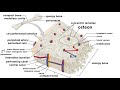 Bones: Structure and Types
 Histology of the cartilage Human anatomy lecture MBBS BDS easy
 Cartilage Science Explained
 Histology of Parotid Gland\ Serous Salivary Gland Histology
 Hyaline cartilage histo diagram
 Histology of Uterus - Secretory phase or Luteal phase
 Hyaline cartilage: slides and function (preview) - Human Histology | Kenhub
 Histology of Cerebral Cortex.
 HISTOLOGY OF THYROID GLAND || Epithelium || Follicular cells || Parafollicular cells || Structure |
 Types of Cartilage | Hyaline, Elastic, and Fibrocartilage
 Histology of Spinal Cord
 Histology Of Elastic Cartilage
 Connective Tissue
 Histology of the Liver
 HISTOLOGY OF TRACHEA || Layers || Hyaline Caritilage
 Histology of Cartilages - Anatomy Videos by Dr. Ashwani Kumar
 Cartilage | Muscular-skeletal system physiology | NCLEX-RN | Khan Academy
 Tetralogy of Fallot (TOF)