Rotator Cuff MRI - Everything You Need To Know - Dr. Nabil Ebraheim
Educational video describing MRI imaging of the rotator cuff.
MRI is valuable in diagnosing most shoulder problems, especially rotator cuff pathology.
Normal rotator cuff anatomy coronal vies: normal cuff appears dark on T1 & T2 MRI. The normal distance between the acromion and the humeral head is between 7-14 mm. in the axial view, check the biceps tendon for subluxation. Subluxation could indicate a subscapularis tendon tear.
Normal rotator cuff sagittal view: normally the supraspinatus muscle occupies the fossa in the sagittal view. When the muscle is abnormal, it does not occupy the fossa. Muscle fat atrophy is usually seen on the sagittal image.
Impingement
Internal impingement is pathology on the underside of the rotator cuff. External impingement (subacromial) is pathology on the bursal side of the rotator cuff and is the most common type of shoulder impingement.
What is internal impingement? internal impingement is partial tear of the under surface of the rotator cuff that occurs in throwing athletes and usually is associated with posterior and superior labral tears.
What is external impingement (sub acromial)? External impingement involves the sub acromial bursa and with external impingement, the pain is increased by overhead activity. The patient could have night pain. The distance between the acromion and the humeral head is usually 6 mm or less. May be associated with type III hooked acromion and Os acromial.
Treatment of sub acromial impingement
•Therapy for at least 6 months before surgery.
Tendinopathy
Rotator cuff becomes thickened with intermediate signal on T1 and T2.
Calcific tenditis
Calcification inside the rotator cuff tendon.
Rotator cuff tears:
Partial three types: fluid or dye extends partially through the thickness of the tendon.
1-Articular surface partial
2-Bursal surface partial
3-Intrasusbtance partial
Full thickness tear: fluid bright signal extends completely through the tendon from superior to inferior. The tendon may be retracted and there will be a gap in the tendon.
MRI arthrogram can improve visualization of the tear and accuracy of the study in asymptomatic patients 60 years old or older, 55% will have a rotator cuff tear on their MRI. If you see a cysts in the humeral head on MRI, the patient will probably have a rotator cuff tear.
Massive tears of the rotator cuff that are greater than 5 cm usually involve multiple tendons.
MRI will show massive cuff tear with retraction at the level of the glenoid with atrophy of the muscle and fatty infiltration. The supraspinatus is ruptured and retracted.
Become a friend on facebook:
http://www.facebook.com/drebraheim
Follow me on twitter:
https://twitter.com/#!/DrEbraheim_UTMC
Видео Rotator Cuff MRI - Everything You Need To Know - Dr. Nabil Ebraheim канала nabil ebraheim
Показать
Комментарии отсутствуют
Информация о видео
Другие видео канала
 Rotator Cuff Muscles - Everything You Need To Know - Dr. Nabil Ebraheim
 Best Way To Know If You Have a Rotator Cuff Tear?
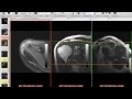 Systematic Interpretation of Shoulder MRI: How I do it
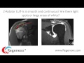 How to read your shoulder MRI with Dr. Centeno of Regenexx
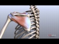 Shoulder Anatomy Animated Tutorial
 Rotator Cuff Tear ,injury - Everything You Need To Know - Dr. Nabil Ebraheim
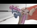 Rotator Cuff Repair with Arthrex® SpeedBridge™
 Shoulder Impingement Syndrome - Everything You Need To Know - Dr. Nabil Ebraheim
 The Exam for Shoulder Pain - Stanford Medicine 25
 Rotator Cuff Tears: What Is the Evidence? - Brian Feeley, MD
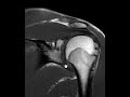 How to read an MRI of the shoulder
 Shoulder Impingement - Dr. Richard Hawkins
 Rotator Cuff | 3D Anatomy Tutorial
 Shoulder Examination Inspection & Palpation - Everything You Need To Know - Dr. Nabil Ebraheim
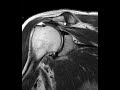 Rotator cuff tear
 Rotator Cuff Tears and Rehabilitation
 Interpretation of Shoulder MRI: Detailed Anatomy
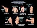 Shoulder Examination / Subacromial, Cuff - Everything You Need To Know - Dr. Nabil Ebraheim
 Rotator Cuff Tears: Do You Need Surgery?
 Top 3 Signs Of A Rotator Cuff Tear (Updated)