Gracilis Muscle Anatomy - Everything You Need To Know - Dr. Nabil Ebraheim
Dr. Ebraheim’s educational animated video teaches the anatomy of the Gracilis muscle with simple images, this video also provides you with all you need to know about this muscle, its innervation, action, and function.
Gracilis is the most superficial muscle on the medial side of the thigh.
Its tendon can be easily palpable in the inguinal region, together with the adductor longus muscle.
The medial side of the thigh contains the adductor group of muscles which help in crossing of the legs and balances the pelvis while walking and standing.
Gracilis is the only adductor muscle that crosses 2 joints.
This thin long muscle provides a reliable coverage.
It is one of the most common donor muscles for free muscle transfer procedures.
Origin: from the outer surface of the ischiopubic ramus.
Insertion: in the upper medial part of the tibial shaft below the sartorious muscle.
This area is called the pis anserine, and will have the Gracilis, Sartorius, and the Semitendinosis.
Innervation: the anterior branch of the obturator nerve.
The Oturator nerve arises from the lumbar plexus L2-L3-L4.
Then the nerve passes through the obturator foramen to reach the adductor muscles.
Function: it adducts the hip, flexes and internally rotates the knee.
Become a friend on facebook:
http://www.facebook.com/drebraheim
Follow me on twitter:
https://twitter.com/#!/DrEbraheim_UTMC
Donate to the University of Toledo Foundation Department of Orthopaedic Surgery Endowed Chair Fund:
https://www.utfoundation.org/foundation/home/Give_Online.aspx?sig=29
Видео Gracilis Muscle Anatomy - Everything You Need To Know - Dr. Nabil Ebraheim канала nabil ebraheim
Показать
Комментарии отсутствуют
Информация о видео
Другие видео канала
 Anatomy Of The Sartorius Muscle - Everything You Need To Know - Dr. Nabil Ebraheim
 Best Self-Treatment for a Groin Pull- Including Stretches & Exercises.
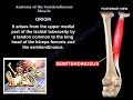 Anatomy Of The Semitendinosus Muscle - Everything You Need To Know - Dr. Nabil Ebraheim
 Anatomy Of The Adductor Magnus Muscle - Everything You Need To Know - Dr. Nabil Ebraheim
 Anatomy Of The Gastrocnemius Muscle - Everything You Need To Know - Dr. Nabil Ebraheim
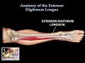 Anatomy Of The Extensor Digitorum Longus Muscle - Everything You Need To Know - Dr. Nabil Ebraheim
 MUSCULAR SYSTEM ANATOMY: Medial thigh region muscles model description. Somso
 Anatomy Of The Sartorius Muscle - Everything You Need To Know - Dr. Nabil Ebraheim
 Gracilis Muscle 3D Animation 4K
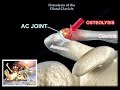 Distal Clavicle Osteolysis - Everything You Need To Know - Dr. Nabil Ebraheim
 Phelps Test | Gracilis Contractures
 Yoga For Psoas | Yoga With Adriene
 Functions of the Sartorius Muscle (preview) - 3D Human Anatomy | Kenhub
 Humeral Shaft Fracture Treatment - Everything You Need To Know - Dr. Nabil Ebraheim
 Gracilis Muscle - Function & Origins - Human Anatomy | Kenhub
 Slipped Capital Femoral Epiphysis - Everything You Need To Know - Dr. Nabil Ebraheim
 Femoral Shaft Fracture in Children - Everything You Need To Know - Dr. Nabil Ebraheim
 Sartorius Muscle - Origin, Insertion, Innervation & Actions - Anatomy | Kenhub
 Anatomy Of The Pectineus Muscle - Everything You Need To Know - Dr. Nabil Ebraheim
 Gracilis And Knee Pain | WODdoc | P365 | Episode 1319