Blood supply and Nerve supply of Long bone - General Anatomy animations
Join this channel to get access to perks:
https://www.youtube.com/channel/UCG5TBPANNSiKf1Dp-R5Dibg/join
►𝐃𝐨𝐰𝐧𝐥𝐨𝐚𝐝 𝐭𝐡𝐞 𝐌𝐞𝐝𝐯𝐢𝐳𝐳 𝐚𝐩𝐩 𝐮𝐬𝐢𝐧𝐠 𝐭𝐡𝐞 𝐛𝐞𝐥𝐨𝐰 𝐥𝐢𝐧𝐤 👇👇👇👇 𝐃𝐨𝐰𝐧𝐥𝐨𝐚𝐝 👇👇👇👇
►𝐀𝐧𝐝𝐫𝐨𝐢𝐝 :- https://bit.ly/3ansFKq
►𝐉𝐨𝐢𝐧 𝐓𝐡𝐢𝐬 𝐂𝐡𝐚𝐧𝐧𝐞𝐥 𝐓𝐨 𝐆𝐞𝐭 𝐀𝐜𝐜𝐞𝐬𝐬 𝐓𝐨 𝐏𝐞𝐫𝐤𝐬 :- https://bit.ly/2RQHvTN
📌𝐅𝐨𝐥𝐥𝐨𝐰 𝐨𝐧 𝐈𝐧𝐬𝐭𝐚𝐠𝐫𝐚𝐦 :- https://www.instagram.com/drgbhanuprakash
Blood supply and Nerve supply of Long bone - General Anatomy animations
Blood is supplied to mature compact bone through the Haversian canal. Haversian canals are formed when individual lamellae form concentric rings around larger longitudinal canals (approx. 50 µm in diameter) within the bone tissue. Haversian canals typically run parallel to the surface and along the long axis of the bone. The canals and the surrounding lamellae (8–15) are called a Haversian system or an osteon. A Haversian canal generally contains one or two capillaries and nerve fibers. The Haversian canals also surround nerve cells throughout the bone and communicate with osteocytes in lacunae (spaces within the dense bone matrix that contain the living bone cells) through canaliculi. This unique arrangement is conducive to the storage of mineral salt deposits that give bone tissue its strength.
The vascular supply of long bones depends on several points of inflow, which feed complex sinusoidal networks within the bone. These in turn drain to various channels through all surfaces of the bone except that covered by articular cartilage.
Volkmann’s canals are channels that assist with blood and nerve supply from the periosteum to the Haversian canal. One or two main diaphyseal nutrient arteries enter the shaft obliquely through one or two nutrient foramina leading to nutrient canals. Their sites of entry and angulation are almost constant and characteristically directed away from the growing epiphysis.
Except for a few with double or no foramina, 90% of long bones have a single nutrient foramen in the middle third of the shaft. The nutrient arteries divide into ascending and descending branches in the medullary cavity. These approach the epiphysis dividing into smaller rami. Near the epiphysis, they anastomose with the metaphyseal and epiphyseal arteries.
The blood supply of the immature bones is similar, but the epiphysis is a discrete vascular zone separated from the metaphysis by the growth plate. Epiphyseal and metaphyseal arteries enter on both sides of the growth cartilage, with anastamoses between them being few or absent.
Growth cartilage receives its blood supply from both sources and also from an anastamotic collar in the adjoining perichondrium. Young periosteum is more vascular, has more metaphyseal branches, and its vessels communicate more freely with those of the shaft than adult periosteum.
Key Points
Haversian canals typically run parallel to the surface and along the long axis of the bone and generally contain one or two capillaries and nerve fibers.
Volkmann’s canals are channels that assist with blood and nerve supply from the periosteum to the Haversian canal.
The vascular supply of long bones depends on several points of inflow.
Except for a few with double or no foramina (places in bone where capillaries enervate), 90% of long bones have a single nutrient foramen in the middle third of the shaft.
Young periosteum is more vascular and its vessels communicate more freely with those of the shaft compared to adult periosteum.
#bloodsupplyoflongbone #nervesupplyoflongbone #longbone #generalanatomy #arterialsupplyoflongbone #longbone #anatomy #nationalexittest #usmle #usmlestep1 #mbbs #plab #neetpg #nationalexittest #medvizz #drbhanuprakash
Видео Blood supply and Nerve supply of Long bone - General Anatomy animations канала Dr.G Bhanu Prakash Animated Medical Videos
Показать
Комментарии отсутствуют
Информация о видео
29 августа 2021 г. 17:30:15
00:04:53
Другие видео канала
 Blood Supply of Long Bone | ANIMATION | USMLE | Anatomy, Lecture | NEET PG, MRCS, The Young Orthopod
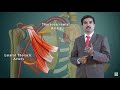 AXILLARY ARTERY ANATOMY ANIMATED LECTURE
 General Anatomy Types of Epiphysis | Mnemonic & Explanation | Anatomy
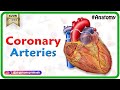 Coronary arteries Anatomy / Blood supply of Heart / Arterial supply of heart : Animation
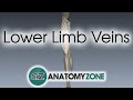 Lower Limb Veins Overview - 3D Anatomy Tutorial
 Structure Of Bone Tissue - Bone Structure Anatomy - Components Of Bones
 SKELETON BONES SONG - LEARN IN 3 MINUTES!!!
 YOUNG LONG BONE || Blood Supply || Long Bone Anatomy || Drawn & Explained
 Long Bone Anatomy - Drawn & Defined
 Femoral Nerve Anatomy: Origin, Course, Branches and Clinical application
 The 6 Types of Joints - Human Anatomy for Artists
 Bone remodeling and repair
 Clavicle Bone Anatomy: Bony Landmarks and Articulations, Functions, Attachments, Clinical aspects
 Radial nerve Anatomy USMLE- Origin, Course, innervation, Saturday night palsy, Wartenberg’s syndrome
 BLOOD SUPPLY OF LONG BONE | Types of Bone| Shape | Arterial Supply | Venous Drainage | Clinical Anat
 Bones: Structure and Types
 The Skeletal System
 Intramembranous Ossification
 MSK Skeletal System Basics - Bone Formation
 Blood Supply of Femoral Head | MADE EASY | NEET PG | Femur Anatomy - The Young Orthopod