MUSCLES OF FACIAL EXPRESSION AND MASTICATION
3D model from https://www.turbosquid.com/3d-models/realistic-head-muscles-anatomy-max/621709
There are several groups of facial muscles: the oral group – in other words, muscles surrounding the mouth, the orbital group – or muscles associated with the eye socket, the nasal group – muscles surrounding the nose, the auricular group – or muscles surrounding the ears, and finally, muscles of the forehead and neck.
Let’s start with the oral group. Most of the facial muscles are positioned around the mouth, since this is the part of the face that moves the most. Firstly, there’s the orbicularis oris, which encircles the mouth. It is a sphincter – aka a circular muscle that maintains constriction of an orifice or passage of the body. This muscle helps you close your mouth, or pucker up your lips. It originates from the maxilla and from the other muscles of the cheek and inserts into the lips. The other muscles from the oral group can be divided into an upper and lower group. The upper group includes the zygomaticus major and minor, the risorius, the levator labii superioris, the levator labii superioris alaeque nasi, and the levator anguli oris.
The zygomaticus major and minor help lift the corners of your lips so you can smile. On the other hand, the risorius muscle is involved in the “fake smile”. Its contraction draws back the corners of the mouth into a smile shape. However, this smile does not collaborate with the orbicularis oculi muscles and so does not involve the skin around the eyes.
The levator anguli oris also plays a role in helping you smile by moving the corners of the mouth upwards. Meanwhile, the levator labii superioris alaeque nasi enables a snarling expression.
The lower group of oral muscles include the mentalis, the depressor anguli oris, and the depressor labii inferioris. The mentalis muscle is located at the furrow between your lower lip and chin, and is nicknamed the pouting muscle, since it helps contract the chin when expressing displeasure. The depressor anguli oris pulls down the corners of your mouth. Meanwhile, the depressor labii inferioris draws the lower lip down and sideways. So in general, the upper group generally is more involved in happy expressions, and the lower group is more involved in upset expressions.
The orbital group includes the orbicularis oculi, the corrugator supercilia, and the depressor supercilia. So the orbicularis oris isn’t the only muscle shaped like a donut. You also have a pair of orbicularis oris muscles encircling your eyelids. These muscles help you close your eyes, and again, these are sphincter muscles. The corrugator muscle pulls the eyebrows medially in most people. Finally, there is the depressor supercilia, which are thought to assist in moving and lowering the eyebrows.
The nasal group has three muscles in it. The nasalis is the largest, and can be split into the transverse nasalis and dilator naris. The transverse naris compresses the nares – or nostrils, while the dilator naris opens them. The procerus originates from the nasal bone, and inserts into the lower medial forehead. Its contraction pulls the eyebrows downward, wrinkling the nose. Finally, there’s the depressor septi nasi, which assists the alar part of the nasali in opening the nostrils. It runs from the maxilla to the nasal septum – the bone and cartilage separating the nasal cavity into two nostrils. There are three auricular muscles – the anterior, posterior, and superior auricular muscles.
Finally there is a muscle of facial expression in the forehead, and one in the neck. The frontalis muscle raises the eyebrows and wrinkles the forehead. In the neck, we have the platysma. It has three portions – the nodular, labial, and mandibular. Activation of the platysma causes slight wrinkling of the skin overtop.
Now for the muscles of mastication – these are associated with movements of the jaw. There are four of these – the temporalis, the masseter, and the medial and lateral pterygoids. The temporalis muscle closes the mouth and retracts the mandible. It originates from the temporal fossa and condenses into a tendon which connects to the mandible. The masseter is the strongest of the muscles of mastication. It can be split into two parts – the superficial masseter, and the deep masseter. Both these muscles elevate the mandible to close the mouth.
The medial pterygoid muscle closes the jaw by elevating the mandible. It has two heads – a deep one and a superficial one. The lateral pterygoid again has two heads – a superior and inferior one. Both heads are involved during the opening of the mouth – basically making sure the skull and jawbone are aligned properly at the temporomandibular joint. Finally, there’s the buccinator! This is an accessory muscle of mastication. Located between the mandible and maxilla, this muscle pulls the cheek in towards the teeth so food doesn’t accumulate there.
Видео MUSCLES OF FACIAL EXPRESSION AND MASTICATION канала Neural Academy
Показать
Комментарии отсутствуют
Информация о видео
Другие видео канала
 Muscles of facial expression (anatomy)
 Cranial Nerve BASICS - The 12 cranial nerves and how to REMEMBER them!
 Skull bones, sutures and landmarks
 THE MUSCLES SONG (Learn in 3 Minutes!)
 Muscles of the neck
 BONES OF THE SKULL - LEARN IN 4 MINUTES
 Facial nerve & facial expression (anatomy)
 Temporomandibular joint & muscles of mastication
 Circulatory System | Arteries & Veins of the Head & Neck | Head Model
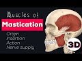 MUSCLES OF MASTICATION - 3D
 Muscles of facial expression
 Muscles of mastication made easy!
 Muscles of the Head & Neck | Anatomy Model
 Good and Bad Facial Muscles for a Beautiful Smile | How to relax bad muscles and activate good ones
![Muscles of Mastication [Part 1] | Origins, Insertions, Etc.](https://i.ytimg.com/vi/gCl_C64od_E/default.jpg) Muscles of Mastication [Part 1] | Origins, Insertions, Etc.
Muscles of Mastication [Part 1] | Origins, Insertions, Etc. Destroying Flat Earth Without Using Science - Part 3: Airplanes
 MUSCLES OF FACIAL EXPRESSION AND MASTICATION SONG
 Muscles of mastication (preview) - Origin, insertion, functions - Human Anatomy | Kenhub
 Extraocular Muscles | Eye Anatomy
 Sensory nerves of the face (trigeminal nerve, CN V anatomy)