SDS PAGE II Protein Electrophoresis- Sodium Dodecyl Sulfate Polyacrylamide Gel Electrophoresis
This video contains
1- SDS PAGE sample preparation
2-Significance of SDS/ Role of SDS
3-Significance of 2-mercaptoethanol/ role of 2-mercaptoethanol
4- Significance of Glycerol/ Role of Glycerol
5-Significance of tracking dye/ Role of Tracking dye
6-Significance of TEMED/ Role of TEMED
7-Significance of APS/ Role of APS
8-Significance of acrylamide/ Role of acrylamide
9-Significance of bis-acrylamide/ Role of bis-acrylamide
10- SDS PAGE gel preparation
11- Polymerization of SDS PAGE gel
12-Principle of SDS PAGE
13- Detection of proteins using coomassie brilliant blue/ silver staining
14- Application of SDS PAGE
SDS–polyacrylamide gel electrophoresis (SDS–PAGE) is the most widely used method for analysing protein mixtures qualitatively. It is particularly useful for monitoring protein purification
Samples to be run on SDS–PAGE are firstly boiled for 5 min in sample buffer containing b-mercaptoethanol and SDS. The mercaptoethanol reduces any disulphide bridges present that are holding together the protein tertiary structure, and the SDS binds strongly to, and denatures, the protein. Each protein in the mixture is therefore fully denatured by this treatment and opens up into a rod-shaped structure with a series of negatively charged SDS molecules along the polypeptide chain. On average, one SDS molecule binds for every two amino acid residues. The original native charge on the molecule is therefore completely swamped by the negatively charged SDS molecules
The sample buffer also contains an ionisable tracking dye, usually bromophenol blue, that allows the electrophoretic run to be monitored, and sucrose or glycerol, which gives the sample solution density thus allowing the sample to settle easily through the electrophoresis buffer to the bottom when injected into the loading well
Once the samples are all loaded, a current is passed through the gel and the proteins are separated.
Video transcript:
SDS-PAGE is mainly carried out for the separation of proteins. For carrying out SDS-PAGE, the sample, i.e. the protein samples are prepared using tris-buffer which maintains the pH, glycerol which gives the sample enough density so it settles inside the well.
Bromophenol blue which is a tracking dye, 2-mercaptoethanol and SDS.
A protein molecule exists in a folded secondary or tertiary structure in its native form.
In these structure disulfide bonds are formed between the sulfur residues on two cysteine molecules.
b-mercaptoethanol basically reduces these di-sulfide bonds, leading to a denatured unfolded form of protein. this, denaturation is triggered by heat.
SDS binds to the unfolded protein and solubilizes it. SDS also provides an overall negative charge to the entire protein. Approximately 1 SDS molecule binds to 2 amino
acids. Proteins differ from each other on the basis
of size as well as charge.
But since the proteins are denatured with an overall negative charge due to SDS. the movement on the gel will not be based on the charge of the protein, but only on the size of the protein.
the concentration of the gel depends on the amount of acrylamide added in the buffer.
Thus, the pore size of the gel, depends upon the concentration of acrylamide that is used
to prepare the gel.
Saturated butanol, or isopropanol is added to avoid oxygen and to facilitate proper polymerization of the gel matrix.
A polyacrylamide gel is made up of acrylamide and bis-acrylamide monomers which are polymerized to form the gel matrix. the polymerization is done using TEMED and
APS. TEMED decomposes Ammonium persulfate to give
free radicals. Free radicals species are highly reactive, they have an unpaired electron, and thus they form single bond with another monomers. this creates an equally reactive radical which
will react with another monomer and form long chains.
The long chains are cross-linked by bis-acrylamide.
The stacking gel has low acrylamide concentration, i.e. it has a high pore size that allows the proteins to migrate freely and get stacked between the interface of the stacking gel and the running gel.
The resolving gel contains a high concentration of acrylamide, i.e. it is having less pore size, which is capable of retarding the movement of proteins.
Thus, it is the resolving gel where the separation of protein molecules takes place.
Another difference between the stacking gel
and the resolving gel lies in the pH.
Stacking gel pH= 6.8
Resolving gel pH= 8.8
Running buffer pH= 8.3
Glycine can exist in three different charged staged, i.e. positive, neutral, or negative depending on the pH.
Low pH= positive charge
High pH= negative charge
Neutral pH- zwitter ion
The proteins move in this sandwich manner till it hits the running gel.
After the gel is run, it can be removed and stained using coomasie brilliant blue, or
silver nitrate to visualize the protein.
Western blot can also be carried out with the gel.
Видео SDS PAGE II Protein Electrophoresis- Sodium Dodecyl Sulfate Polyacrylamide Gel Electrophoresis канала BioMagica
Показать
Комментарии отсутствуют
Информация о видео
Другие видео канала
 blood typing- ABO blood grouping & co dominance
 Pedigree Identification Quick Tips
 Codominance II Non-mendelian inheritance
 Ecogeographical rules II Ecology Unit II CSIR NET II Ecology Rules
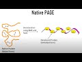 Native PAGE : Polyacrylamide gel electrophoresis II Protein Electrophoresis
 DNA structure Mneumonic-Purine and Pyrimidines structures made easy
 Polyacrylamide Gel Electrophoresis II Protein Electrophoresis Basics II PAGE Basics
 CSIR NET Life Sciences- UNIT 2 part B II 2012
 Excision Repair of DNA (Nucleotide Excision Repair and Base Excision repair)
 Principle of Blood Agar medium II Microbiology II Media principle
 2D polyacrylamide gel electrophoresis : 2D PAGE
 Principle of Coagulase test II Biochemical test II Microbiology
 Luria Delbruck Experiment (Fluctuation Test) II DNA mutations and repair mechanisms
 Detect apoptosis using Flow cytometry analysis- CSIR NET
 Two point Mapping II 3 Easy Steps II Recombination frequency II Linkage
 Tips & Tricks for Pedigree analysis
 Principle of Salt Mannitol Agar-Isolation of S.aureus (Staphylococcus isolation)
 Principle of Xylose lysine deoxycholate agar (XLD agar) II Microbiology II Media principle
 Types of Point Mutation II Transversion, Transition ( Neutral, silent and missense mutation)
 Principle of MacConkey’s Agar II Microbiology II Media principle