Mitosis in Onion Root tip Experiment
Real photographs of all the Mitotic Stages taken by me. Check out my Genetics experiment videos. Go to my Channel page, click "Videos". Do show ur support, SUBSCRIBE!
Also do click on the HASHTAGS above this Video title to watch my other videos on genetics experiments and many more!
#GeneticsLab #CytologyLab #ThomasTKtungnung
Mitosis in Onion Root tip Experiment
In this video, we’ll be preparing root tips of Allium species to observe cells in various stages of mitotic division
Mitosis is one of the stages in the cell cycle where the chromosomes of a cell replicate and separate to eventually form two genetically identical cells. Unlike in meiosis, chromosome number is maintained in both daughter cells.
Active mitosis in Allium species occurs twice in a 24hour period with a primary maximum phase at around 11pm and a secondary maximum phase at around 1pm.
So based on this information, it goes without saying that these two time periods would be the most preferable for collecting root tip for mitosis lab work.
For the experiment, you’ll need:
• Onion or Garlic bulb
• Beakers
• Toothpicks
• Carnoy’s fluid
• 70% ethanol
• 1N Hydrochloric acid
• Aceto-carmine or Aceto-orcein stain
• Glass slides and coverslips
• Blade or scalpel
• Watchglasses or petridishes
• Tiny vials or containers
• Spirit lamp or Bunsen burner or any flame source
• Blotting paper
• Droppers
• Thumb forceps and a pair of scissor
• Compound light microscope and
• Immersion oil
To begin the experiment, take a onion or garlic bulb and fix in on a beaker containing tap water using toothpicks like you see here. Make sure the base of the bulb touches the water level. Keep this in a safe corner for a couple of days.
Once the roots have grown about 2-3cms, cut out 1cm of root tips and transfer them into a tiny vial containing carnoys fixative fluid. Carnoy’s fluid fixes the DNA of the root tip cells. Leave the roots in the fixative for about 48hrs.
If your planning on storing the root tips for a longer time for use in future, you may transfer the fixed root tips in a second vial containing 70% ethanol. Ethanol dehydrates the root tissue and thereby preserves DNA. Root tips preserved in ethanol and preferably refrigerated will keep well for a couple of years for genetic studies.
Either ways, take a few root tips from carnoy fluid or ethanol and transfer them onto a watchglass containing 1N Hydrochloric acid. Acid will soften the cell walls and weaken cellular connections so it becomes easy to squash the root material later.
Gently warm the watchglass on a flame for about 5 secs. Expose the roottips in the acid for about 2mins.
Give the root tips a couple of washings in distilled water.
Now transfer the roots onto a watchglass containing acetocarmine or acetoorcein stain. These are excellent chromosomal stains and will impart a deep red colouration to the nuclear material of the root cells.
Warm the stain on the flame for about 5secs and leave the roottips in the stain for about 5-10mins.
Now transfer the roottips onto a clean glass slide containing a drop of water.
Using a sharp blade or scalpel, remove about a millimetre of the root tips and discard the rest. The very tip s of the roots are the regions with active cell division and they are all that we require for mitotic experiments.
Gently lower a coverslip on the root tips making sure to avoid any air bubbles.
Using the blunt end of a forcep or a pencil, gently tap the coverslip a few times until the root tips are uniformly squashed in between the slide and the coverslip.
A properly squashed slide will appear faint cloudy pink to almost colourless.
The roottip slide is now ready for microscopic observation.
This is how a finely squashed roottip appears under 10x of the microscope. As you can see, the cells of the roottips are nicely spread out with almost not overlappings. If root tips are not squashed enough, you may repeat the squashing process until you get well spread root cells.
Scan the slide for cells showing mitotic division stages under low power and then proceed to observation under high power, such as 40x or 100x oil immersion lens.
For observations under 100x objective, you”ll neeed to add a drop of immersion oil on top of the coverslip and gently lower the objective lens until it touches the oil surface. From there, use the fine adjustment knob for focusing.
You may take microphotographs of the various cell division stages by employing a simple technique using a mobile phone and a tripod, like so.
Видео Mitosis in Onion Root tip Experiment канала ThomasTKtungnung
Показать
Комментарии отсутствуют
Информация о видео
Другие видео канала
 See a Salamander Grow From a Single Cell in this Incredible Time-lapse | Short Film Showcase
 mitosis 3d animation |Phases of mitosis|cell division
 🔬 Make Onion mitosis CHROMOSOMES visible - Staining Tutorial | Amateur Microscopy
 Mitosis: The Amazing Cell Process that Uses Division to Multiply! (Updated)
 Amazing Microscopic World! Common Objects Under The Microscope || HOME EXPERIMENTS
 Phases of Mitosis
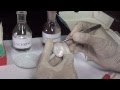 Onion and Cheek Cells - MeitY OLabs
 Total RBC Count Practical Lab
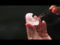 How to Prepare Stained Temporary Mount of Onion Peel | Onion PEEL under microscope (HINDI)
 Mitosis vs. Meiosis: Side by Side Comparison
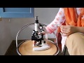 Mitosis slide preparation from onion root tip cells.
 Mitotic Index Root Tip Squash
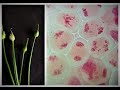 Meiosis in onion flowerbuds experiment
 Differences between Mitosis and Meiosis | Don't Memorise
 Human Sleep Experiment That Went Horribly Wrong
 MEIOSIS - MADE SUPER EASY - ANIMATION
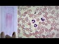 Blood Smear Preparation and Staining Practical Lab
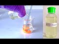 Nessler's Reagent Preparation
 Observation of Mitosis in Onion Root tip Experiment | Practical, Procedure
 ONION ROOT TIP MITOSIS EXPERIMENT