Blood Smear Preparation and Staining Practical Lab
Check out my other Haematology videos! Go to my Channel Page, click on "Videos". Show ur Support, SUBSCRIBE! Do click on the HASHTAGS above this video title to watch my other Haematology experiments and many more in my Playlists!
Total WBC Count: https://www.youtube.com/watch?v=xyhbIPSLBsA&t=364s
Total RBC Count: https://www.youtube.com/watch?v=0f9p9JX4qJk&t=130s
DLC: https://www.youtube.com/watch?v=VFKm_kMTf50
Haemocytometer Use: https://www.youtube.com/watch?v=7IpE33WPRrc&t=43s
Haemometer Hb estimation: https://www.youtube.com/watch?v=6CqptdZyUaU&t=45s
Blood Smear Prep and Staining: https://www.youtube.com/watch?v=KSs0SMfERuA&t=38s
RBCs in iso, hypo and hypertonic solns: https://www.youtube.com/watch?v=urYJAPGUpuw
#HaematologyLab #MedicalLab #ThomasTKTungnung #CytologyLab #AnimalPhysioLab
Thin Peripheral Blood smear preparation and Staining
Blood is a type of fluid connective tissue that carries nutrients and oxygen to various cells in the body and at the same time transports metabolic waste away from the same cells.
Vertebrate blood is composed of blood cells suspended in plasma.
Blood cells mainly consists of Red Blood cells or Erythrocytes or RBCs, White Blood cells or Leucocytes or WBS and and Platelet cells. RBCs are the most abundant blood cells. They impart a red colour to the blood.
In this experiment, we will be covering the procedure on how to prepare a thin blood smear and stain it with an appropriate stain for the morphological and quantitative study of the various cellular elements of blood.
For this experiment, we will need the following:
Blood sample
Glass slides
Leishman’s stain or Wright’s Blood stain or Giemsa’s stain
Distilled water
Droppers
Petridish or Coplin jar
Compound light microscope
Immersion oil
Cotton and rubbing alcohol
Blood lancet or a pricking device
Take two glass slides and clean it with 90% alcohol
Disinfect a finger with alcohol and prick with a sterile lancet or a pricking device
Place a tiny droplet of the blood at the centre of one corner of one of the glass slides.
Using the second pre cleaned slide, touch the drop of blood while inclining the slide at a 45 degree angle.
Now gently and briskly move the inclined slide towards the other end of the slide containing the blood drop in order to obtain a smear.
A properly smeared blood appears roughly tongue shaped as seen here.
Allow the smear to air dry for a minute or so.
Place the slide in a petri dish with the blood smear facing up.
Add several drops of the Leishman’s stain to the blood smear and cover the petri dish.
Alternatively, you may fill a coplin jar with Leishman stain and immerse the slide in the stain.
Stain for 1 minute and then add twice the volume of distilled water to the stain.
Allow the water and the stain to properly mix and keep it aside for about 10minutes.
Drain the slide and wash with distilled water.
Proper staining results in a blood smear that is rose pink in colour.
Air dry the slide while keeping it in an inclined position.
The slide is now ready for observation under the microscope.
Observe and study the various blood cells under 40x and 100x objective.
For 100x , place a drop of immersion oil on the slide, gently lower the 100 x objective until it touches the oil surface. Hereon, use the fine adjustment knob for focussing.
Видео Blood Smear Preparation and Staining Practical Lab канала ThomasTKtungnung
Показать
Комментарии отсутствуют
Информация о видео
Другие видео канала
 Identifying Leukocytes
 LEISHMAN STAINING | Blood Smear Staining Technique | Microbiology | Vivek Srinivas | #Bacteriology
 Hemepath & Hematology Board Review: Peripheral Blood Smears with Dr. Jeanette Ramos
 Malaria Thin Smear Preparation
 Hematology- Making a Peripheral Blood Smear
 PERIPHERAL BLOOD SMEAR | HAEMATOLOGY LAB | PHYSIOLOGY PRACTICALS
 GRAM STAINING | Bacterial Staining Technique | Microbiology | Vivek Srinivas | #Bacteriology
 Malaria Thick Smear Preparation
 Urine Analysis
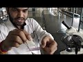 Differential Leukocyte Count By Pandian M. #pvsmededu
 Total RBC Count Practical Lab
 Differential Leucocyte Count DLC Practical Lab
 Blood smear and Giemsa stain
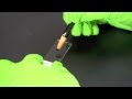 Gram Staining
 hematology in 10 min: Peripheral blood smear examination under microscope
 Total WBC count Practical Lab
 WBC IDENTIFICATION | PERIPHERAL BLOOD SMEAR | HAEMATOLOGY LAB | PHYSIOLOGY
 Part 2 - Examination of Peripheral Blood Smear
 Anatomy Virtual practical exam practice white blood cell slide histology review
 🔬 How to stain cells with Methylene Blue | Amateur Microscopy