How to Prepare Stained Temporary Mount of Onion Peel | Onion PEEL under microscope (HINDI)
How to Download Notes in PDF from Solution Pharmacy Facebook Group Using Laptop
https://youtu.be/cE5MAt0J6hs Using Mobile https://youtu.be/ntzXKi2pA5U
This video is also available in ENGLISH. How to Prepare Stained Temporary Mount of Onion Peel and its Microscopic Study (ENGLISH) https://youtu.be/jQwzmvApC0E
ONION PEEL SLIDE
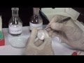
The epidermal cells of onions provide a protective layer against viruses and fungi that may harm the sensitive tissues. Because of their simple structure and transparency, they are often used to introduce students to plant anatomy or to demonstrate plasmolysis. Tissue from an onion is a good first exercise in using the microscope and viewing plant cells. The cells are easily visible under a microscope and the preparation of a thin section is straight forward. An onion is made of layers, each separated by a thin skin or membrane. In this exercise you will make a wet mount on a microscope slide and look at the cells of the onion membrane magnified by the high power, compound microscope.
The epidermis is the protective outer layer of clonally related cells covering all plant organs. It is composed of a number of specialised cell types which differentiate from the basal epidermal cell in adaptively significant frequencies and patterns. The epidermis is unique in developing solely through anticlinal divisions, generating a sheet of cells overlying the rest of the plant. This sheet is connected physically and biochemically to the cell layers below, with information exchange occurring in both directions. The specialised cell types within the epidermis develop either through communication among themselves, or, in some cases, through communication also with the underlying cell layers. Brief descriptions of the molecular genetic control of trichomes, stomata, root hairs and petal conical‐papillate epidermal cells are provided here, along with a summary of the role of the cuticle in epidermal cell morphology and of the interplay between cuticle regulation and cell morphology.
Solution-Pharmacy- The solution-Pharmacy is the completely dedicated channel for Pharmacy Profession. Here we provide Free MCQs, Flashcard and Most Importantly Practical Videos for all Students.
Get in touch with the solution by just clicking following links-
Facebook Group- https://www.facebook.com/groups/solutionpharamcy
Facebook Page- https://www.facebook.com/pharmavideo/
New channel (Pharmacy Dictionary) - https://www.youtube.com/channel/UCt6OXVV_2oxf5DD0Mad6e9A
Instagram- https://www.instagram.com/solutionpharmacy/
E-Mail for official and other work - solutionpharmacy@gmail.com
LinkedIn- http://linkedin.com/in/pushpendrakpatel
#solutionpharmacy #Pharmacologyclass #GPATonlinetest #Pahrmacypracticals
Видео How to Prepare Stained Temporary Mount of Onion Peel | Onion PEEL under microscope (HINDI) канала Solution- Pharmacy
https://youtu.be/cE5MAt0J6hs Using Mobile https://youtu.be/ntzXKi2pA5U
This video is also available in ENGLISH. How to Prepare Stained Temporary Mount of Onion Peel and its Microscopic Study (ENGLISH) https://youtu.be/jQwzmvApC0E
ONION PEEL SLIDE
The epidermal cells of onions provide a protective layer against viruses and fungi that may harm the sensitive tissues. Because of their simple structure and transparency, they are often used to introduce students to plant anatomy or to demonstrate plasmolysis. Tissue from an onion is a good first exercise in using the microscope and viewing plant cells. The cells are easily visible under a microscope and the preparation of a thin section is straight forward. An onion is made of layers, each separated by a thin skin or membrane. In this exercise you will make a wet mount on a microscope slide and look at the cells of the onion membrane magnified by the high power, compound microscope.
The epidermis is the protective outer layer of clonally related cells covering all plant organs. It is composed of a number of specialised cell types which differentiate from the basal epidermal cell in adaptively significant frequencies and patterns. The epidermis is unique in developing solely through anticlinal divisions, generating a sheet of cells overlying the rest of the plant. This sheet is connected physically and biochemically to the cell layers below, with information exchange occurring in both directions. The specialised cell types within the epidermis develop either through communication among themselves, or, in some cases, through communication also with the underlying cell layers. Brief descriptions of the molecular genetic control of trichomes, stomata, root hairs and petal conical‐papillate epidermal cells are provided here, along with a summary of the role of the cuticle in epidermal cell morphology and of the interplay between cuticle regulation and cell morphology.
Solution-Pharmacy- The solution-Pharmacy is the completely dedicated channel for Pharmacy Profession. Here we provide Free MCQs, Flashcard and Most Importantly Practical Videos for all Students.
Get in touch with the solution by just clicking following links-
Facebook Group- https://www.facebook.com/groups/solutionpharamcy
Facebook Page- https://www.facebook.com/pharmavideo/
New channel (Pharmacy Dictionary) - https://www.youtube.com/channel/UCt6OXVV_2oxf5DD0Mad6e9A
Instagram- https://www.instagram.com/solutionpharmacy/
E-Mail for official and other work - solutionpharmacy@gmail.com
LinkedIn- http://linkedin.com/in/pushpendrakpatel
#solutionpharmacy #Pharmacologyclass #GPATonlinetest #Pahrmacypracticals
Видео How to Prepare Stained Temporary Mount of Onion Peel | Onion PEEL under microscope (HINDI) канала Solution- Pharmacy
Показать
Комментарии отсутствуют
Информация о видео
Другие видео канала
 Onion and Cheek Cells - MeitY OLabs
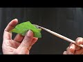 How to Prepare Stomata Slide for Microscopic Study of Stomata (HINDI) By Solution Pharmacy
 11 Fascinating Chemistry Experiments (Compilation)
 Amazing Microscopic World! Common Objects Under The Microscope || HOME EXPERIMENTS
 12 th Biology Practical: study of pollen germination on stigma.
 Onion Cell Microscope Slide Experiment
 How to focus slides under compound microscope // how to use microscope // compound microscope
 To prepare a temporary mount of human cheek epithelial cells, and to study its characteristics
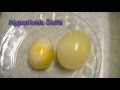 Egg Osmosis (Hypertonic vs. Hypotonic Solution)
 COMMON OBJECTS UNDER MICROSCOPE || 26 HOME EXPERIMENTS
 Study of Plasmolysis - MeitY OLabs
 🔬 097 - How to see moving CELL ORGANELLES of an ONION under the microscope
 Onion and Cheek Cells - MeitY OLabs
 Toxic House Mold Under the Microscope
 My Blood - Zoomed 2000x under the Microscope
 Boiled Dirty Water under the Microscope
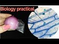 Biology practical| 9th biology practical | prepare slide of onion epidermis
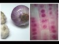 Mitosis in Onion Root tip Experiment
 🔬 098 - How to shrink ONION CELLS - and make them explode as well | citizen science