Neuroanatomy : The Human Brain
For more information : http://neuromatiq.net
For the specific article about the brain : http://neuromatiq.net/en/chapters/2-anatomy/05-the-brain.html
Please : Like, comment, share, subscribe, comment on the web specific page, and you can contribute by either correcting the missing English translations, or translate some articles to a language you master, I can make videos with your sound recordings and credit you for that :).
The human forebrain is made of two hemispheres almost symmetrical, left and right. And a diencephalon, a medial and single part that includes the thalamus and the hypothalamus.
The two cerebral hemispheres are connected by commissural pathways, the biggest is the corpus callosum with more than 200 million fibers crossing from one side to the other.
Each cerebral hemisphere is divided into two distinct regions: a peripheral part, the cortex "gray matter" containing the bodies of nerve cells, and a central part, made of white matter that contains axonal extension of neurons and their myelin sheath.
Each hemisphere is cut by deep fissures that define lobes. The first is the lateral sulcus or Sylvian fissure, where lies the middle cerebral artery, it separates the frontal lobe of the temporal lobe. The second is the central sulcus or fissure of Rolando, between the frontal lobe and the parietal lobe. The third sulcus is the parieto-occipital sulcus, separating the occipital lobe from the temporal and parietal lobes.
In addition there is a fifth lobe, non visible on the surface: the lobe of the insula, found by removing the Sylvian fissure.
In each lobe, there are less deep sulci that delimit the ridges on the cerebral cortex we call gyri.
Видео Neuroanatomy : The Human Brain канала Neuromatiq
Показать
Комментарии отсутствуют
Информация о видео
Другие видео канала
 Neuroanatomy : Diencephalon, Thalamus & Hypothalamus
 Concussion / Traumatic Brain Injury (TBI)
 BRAIN - AN Overview ( For medical/paramedical students only)
 central nervous system || 3d Video|| 3d animation || Biology topic
 Hypothalamus and Limbic System - UBC Neuroanatomy - Season 1 - Ep 4
 Neuroscience Basics: Human Brain Anatomy and Lateralization of Brain Function, 3D Animation.
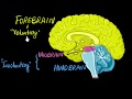 Brain: Parts & functions (Fore, mid & hind) | Control & Coordination | Biology | Khan Academy
 Basal Ganglia 3D Tour
 Neuroanatomy - The Cerebellum
 Neuroanatomy - The Brainstem
 Neuroanatomy: The Cerebrospinal Fluid CSF
 Neuroanatomy - The spinal cord
 The ventricular system
 MRI / 3D Brain Anatomy: Internal Capsule, Thalamus & Brainstem
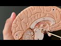 Human Anatomy, Brain Model
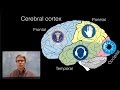 The Brain
 Human Brain Animation | Learn Anatomy Of The Human Brain | iKen | iKen Edu | iKen App
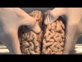 Introduction: Neuroanatomy Video Lab - Brain Dissections
 Human brain and its parts - Biology
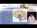 Neuroanatomy made ridiculously simple