The Ventricles: Neuroanatomy Video Lab - Brain Dissections
The ventricles are demonstrated and named on a model cast as well as in rotating 3D reconstructions. The production, function, circulation and removal of CSF produced by the choroid plexus is discussed using a diagram and then reviewed on frontal, axial and sagittal brain specimens and corresponding MRI’s. The blood CSF and brain barriers are mentioned along with the cisterns.
This is 5 of a series of 26 videos to be viewed in the suggested order or intermixed with other curricular materials. The entire series can be accessed here:
https://neurologicexam.med.utah.edu/adult/html/brain-dissections.html
The videos may be downloaded in various formats by going here:
https://neurologicexam.med.utah.edu/adult/html/download_instructions.html
Password Request form for downloadable Neuroanatomy Brain Dissection videos here: https://library.med.utah.edu/neuro-exam/
3D Brain Animations
Neuroanatomy Interactive Syllabus
John Sundsten & Kate Mulligan, University of Washington
Radiological Images
Stephen C. Voron, MD, Associate Professor, Department of Neurobiology & Anatomy, School of Medicine, University of Utah
Specimens
Department of Pathology, School of Medicine, University of Utah
Produced by
Derek Cowan & Suzanne Stansaas, PhD
Copyright 2015, Suzanne Stansaas, PhD, Department of Neurobiology and Anatomy, University of Utah
Видео The Ventricles: Neuroanatomy Video Lab - Brain Dissections канала Eccles Health Sciences Library Digital Publishing
Показать
Комментарии отсутствуют
Информация о видео
7 июля 2015 г. 0:44:06
00:27:20
Другие видео канала
 Basal Ganglia: Neuroanatomy Video Lab - Brain Dissections
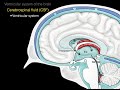 The ventricular system
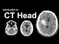 Introduction to CT Head: Approach and Principles
 Circulation in Ventricles and Dural Sinuses
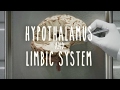 Hypothalamus and Limbic System - UBC Neuroanatomy - Season 1 - Ep 4
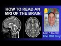 How to Read an MRI of the Brain | First Look MRI
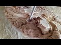 NEUROANATOMY - THIRD VENTRICLE, THALAMUS, HYPOTHALAMUS AND CORPUS CALLOSUM - BY DR MITESH DAVE
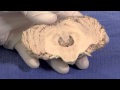 The Cerebellum: Neuroanatomy Video Lab - Brain Dissections
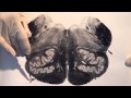 Brain Stem & Reflexes: Neuroanatomy Video Lab - Brain Dissections
 Introduction to Neuroanatomy - Neuroscience - Neurophysiology - Central Nervous System
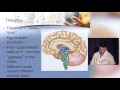 Neuroanatomy made ridiculously simple
 Basal Ganglia - UBC Flexible Neuroanatomy - Season 1 - Ep 7
 Cerebrospinal Fluid - Neuroanatomy
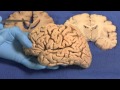 Limbic: Neuroanatomy Video Lab - Brain Dissections
 Introduction to the Central Nervous System - UBC Neuroanatomy Season 1 - Ep 1
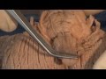 Cranial Nerves: Neuroanatomy Video Lab - Brain Dissections
 Neuroanatomy on MRI | Part 1| Cerebrum, Basal Ganglia, Thalamus, Internal Capsule & Lesions
 Neuroanatomy: The Cerebrospinal Fluid CSF
 Lateral ventricle of brain
 Ventricles of the Brain | Anatomy Model